

Abstract
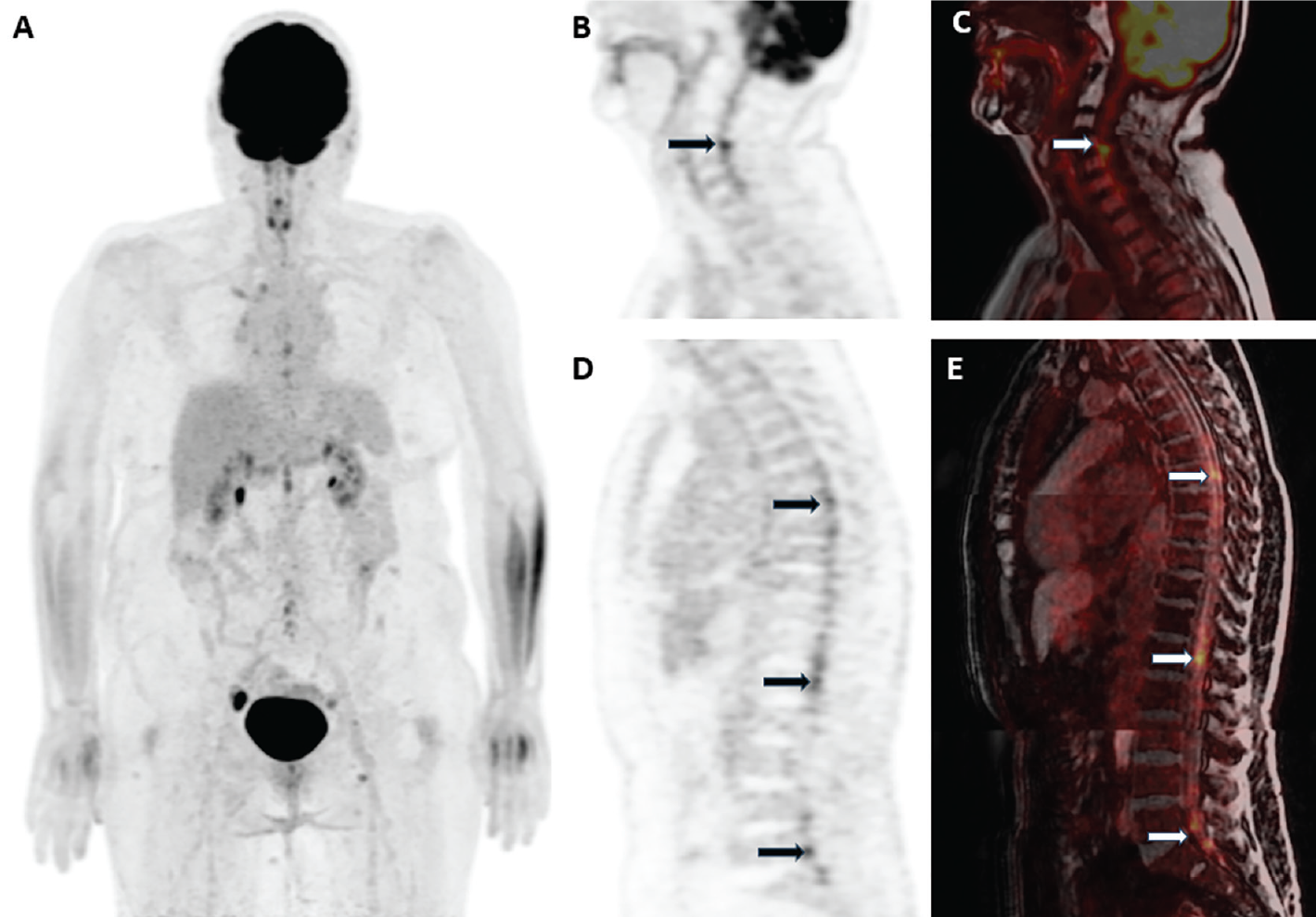
Spinal cord involvement is a rare manifestation of metastatic ovarian carcinoma. We report a case of high-grade serous ovarian cancer initially diagnosed in 2020. After cytoreductive surgery and multiple chemotherapy regimens, the patient developed brain metastases followed by small hypermetabolic foci along the spinal canal detected on 18F-fluorodeoxyglucose positron emission tomography/magnetic resonance imaging (18F-FDG PET/MRI). These lesions, primarily located in the cervical region, were confirmed by contrast-enhanced spinal MRI but were more clearly visualized on 18F-FDG PET/MRI. Intramedullary spinal cord metastases are extremely rare and often underdiagnosed. While MRI remains the standard for anatomical assessment, 18F-FDG PET/MRI demonstrated superior lesion detection and definition in this case. This highlights the potential of hybrid imaging for improved diagnostic sensitivity in patients with suspected central nervous system involvement.
Keywords: 18F-FDG PET/MRI, ovarian carcinoma, spinal cord metastasis, hybrid imaging
,
Central nervous system (CNS) involvement is an uncommon manifestation of metastatic ovarian carcinoma. Though the liver is the most frequent site of distant spread, spinal cord metastases remain rare. The anatomical distribution of metastases is an independent prognostic factor (). We present a case of spinal cord metastasis from high-grade serous ovarian carcinoma, emphasizing the diagnostic value of 18F-FDG PET/MRI compared to conventional MRI.
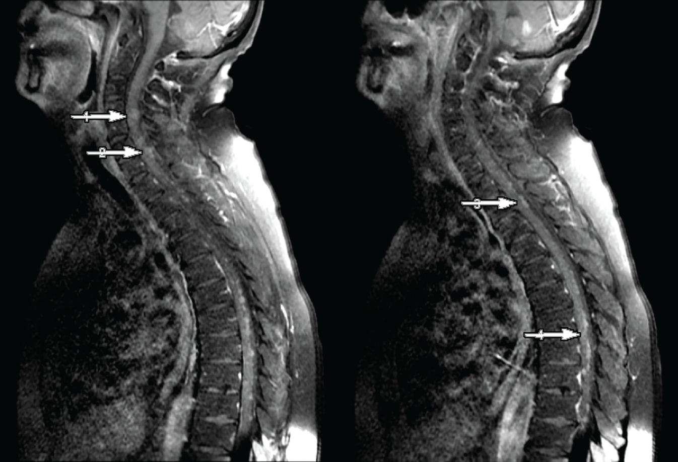
The patient was a 42-year-old female initially diagnosed in 2020 with high-grade serous ovarian carcinoma following cytoreductive surgery that included total hysterectomy, bilateral salpingo-oophorectomy, omentectomy, and excision of multiple peritoneal implants. Based on surgical pathology, the disease was classified as FIGO stage IIIC. Between 2020 and 2024, the patient received multiple lines of systemic chemotherapy, including carboplatin–paclitaxel, gemcitabine, and bevacizumab-based regimens. During follow-up in 2024, brain metastases developed; palliative whole-brain radiotherapy was administered at a total dose of 30 Gy in 10 fractions. Subsequently, 18F-FDG PET/MRI revealed small hypermetabolic foci along the spinal canal particularly in the cervical region adjacent to the spinal cord, suggestive of metastases. Contrast-enhanced spinal MRI confirmed nodular enhancement in the corresponding regions. Contrast-enhanced spinal MRI on post-contrast T1-weighted images demonstrated nodular, contrast-enhancing lesions within the spinal canal along the spinal cord and interspersed between the cauda equina nerve roots, consistent with intraspinal metastatic spread, compatible with leptomeningeal disease. However, compared with the hypermetabolic foci observed on 18F-FDG PET/MRI, precise anatomical delineation of individual lesions was challenging on MRI alone. PET/MRI provided complementary value to spinal MRI by improving lesion detection and delineation through the addition of metabolic imaging. Based on the PET/MRI findings, targeted radiotherapy was initiated.
Spinal cord metastases are extremely rare, occurring in 0.9-2.1% of cancer autopsies and accounting for 8.5% of all CNS metastases. Ovarian cancer cases with spinal involvement are rarely reported and are mostly identified by MRI (-). Nevertheless, several case reports have demonstrated the contribution of 18F-FDG PET/computed tomography to detecting spinal cord metastases in ovarian cancer and other malignancies (-). While MRI remains the gold standard for anatomical evaluation and surgical planning (), 18F-FDG PET/MRI adds diagnostic value, especially for detecting subtle or widespread lesions. As demonstrated in this patient, PET/MRI may represent a diagnostically superior modality in selected clinical contexts.
Footnotes
Ethics
Informed Consent: Informed consent was obtained from the patient for publication.
Authorship Contributions
Concept: C.G., S.A., H.B.S., Design: C.G., S.A., H.B.S., Data Collection or Processing: C.G., Literature Search: C.G., Writing: C.G., S.A., H.B.S.
Conflict of Interest: No conflicts of interest were declared by the authors.
Financial Disclosure: The authors declare that this study has received no financial support.
References
Deng K, Yang C, Tan Q, Song W, Lu M, Zhao W, Lou G, Li Z, Li K, Hou Y. Sites of distant metastases and overall survival in ovarian cancer: a study of 1481 patients. Gynecol Oncol. 2018;150:460-465.
Kłębczyk AW, Wolszczak Z, Urbaniak-Wąsik S, Podlewski S. Metastasis of ovarian cancer to the spinal cord — case study. Oncology in Clinical Practice. 2021;17:81-84.
Miranpuri AS, Rajpal S, Salamat MS, Kuo JS. Upper cervical intramedullary spinal metastasis of ovarian carcinoma: a case report and review of the literature. J Med Case Rep. 2011;5:311.
Safadi S, Rendon P, Rutledge T, Mayasy S. Ovarian carcinoma with isolated spinal cord metastasis. J Investig Med High Impact Case Rep. 2016;4:2324709616657644.
Cormio G, Di Vagno G, Di Fazio F, Loverro G, Selvaggi L. Intramedullary spinal cord metastasis from ovarian carcinoma. Gynecol Oncol. 2001;81:506-508.
Kalimuthu LM, Ora M, Gambhir S. Recurrent renal carcinoma with solitary intramedullary spinal cord metastasis. Indian J Nucl Med. 2020;35:358-9.
Nguyen NC, Sayed MM, Taalab K, Osman MM. Spinal cord metastases from lung cancer: detection with F-18 FDG PET/CT. Clin Nucl Med. 2008;33:356-358.
Krupa M, Byun K. Leptomeningeal carcinomatosis and bilateral internal auditory canal metastases from ovarian carcinoma. Radiol Case Rep. 2017;12:386-390.
Waldron JS, Cha S. Radiographic features of intramedullary spinal cord tumors. Neurosurg Clin N Am. 2006;17:13-19.

