

Abstract
We report a case of a 64-year-old male with metastatic non-small-cell lung carcinoma who was undergoing neoadjuvant chemoimmunotherapy with cisplatin, pemetrexed, and nivolumab. Restaging 18F-fluorodeoxyglucose positron emission tomography/computed tomography (18F-FDG PET/CT) demonstrated segmental, linear 18F-FDG uptake along the abdominal aortic wall with corresponding crescentic soft-tissue thickening. Differential diagnoses included intramural hematoma, focal aortitis, atherosclerotic plaque, and tumor thrombus. CT angiography revealed a mildly enhancing periaortic soft-tissue rim involving a 5 cm infrarenal aortic segment, consistent with isolated periaortitis, likely induced by immunotherapy. Steroid therapy was initiated. Follow-up 18F-FDG PET/CT showed complete metabolic resolution, supporting an immune-related etiology. 18F-FDG PET/CT played a crucial role in early recognition of this immune-related adverse event by sensitively detecting vascular inflammation and guiding further evaluation with CT angiography. Early PET-based detection enabled timely initiation of steroid therapy, preventing potential vascular complications.
Keywords: 18F-FDG PET/CT, immune checkpoint inhibitors, periaortitis, immune-related adverse events, anti-programmed-death-receptor-1 (PD-1) antibody
,
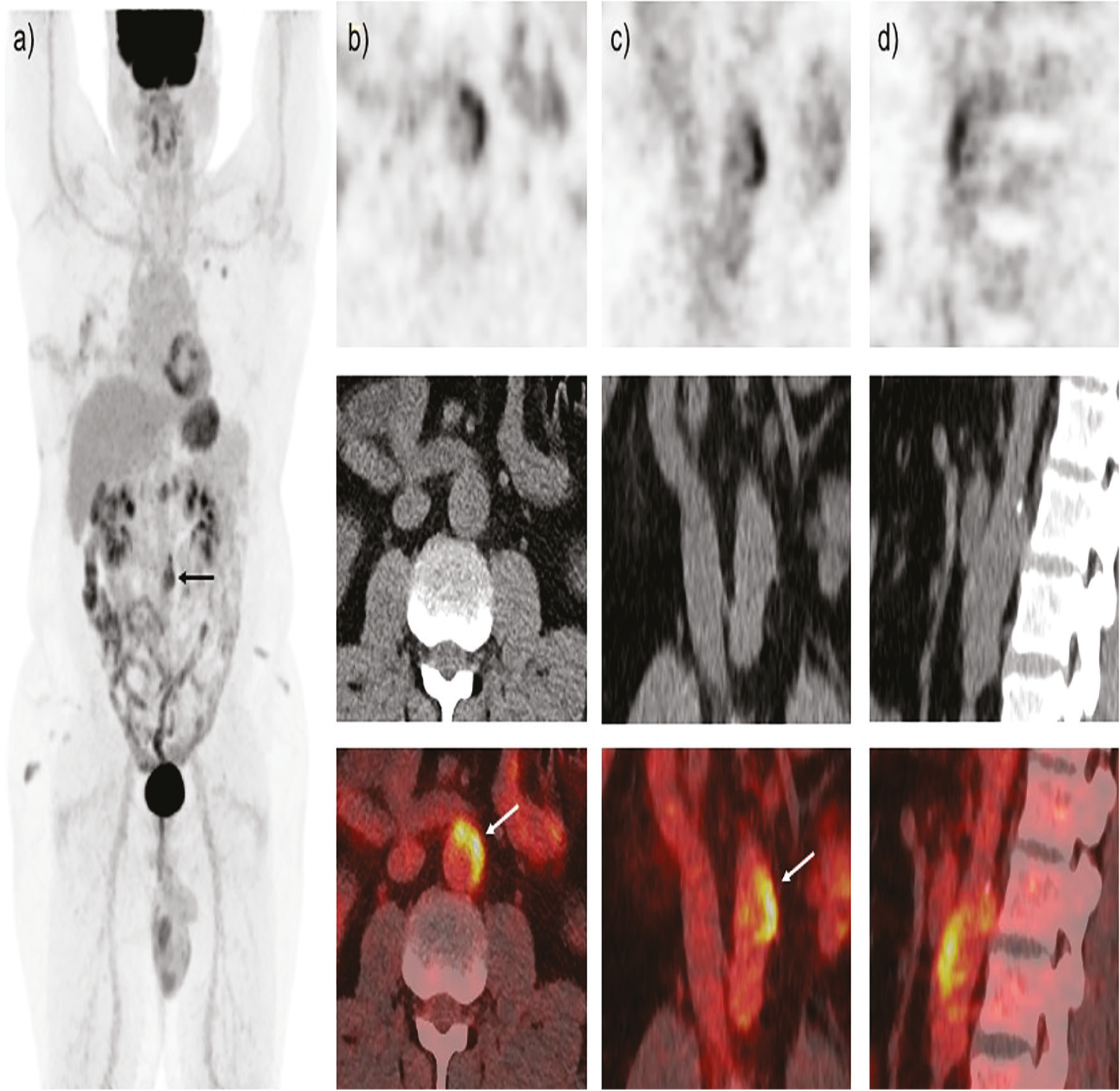
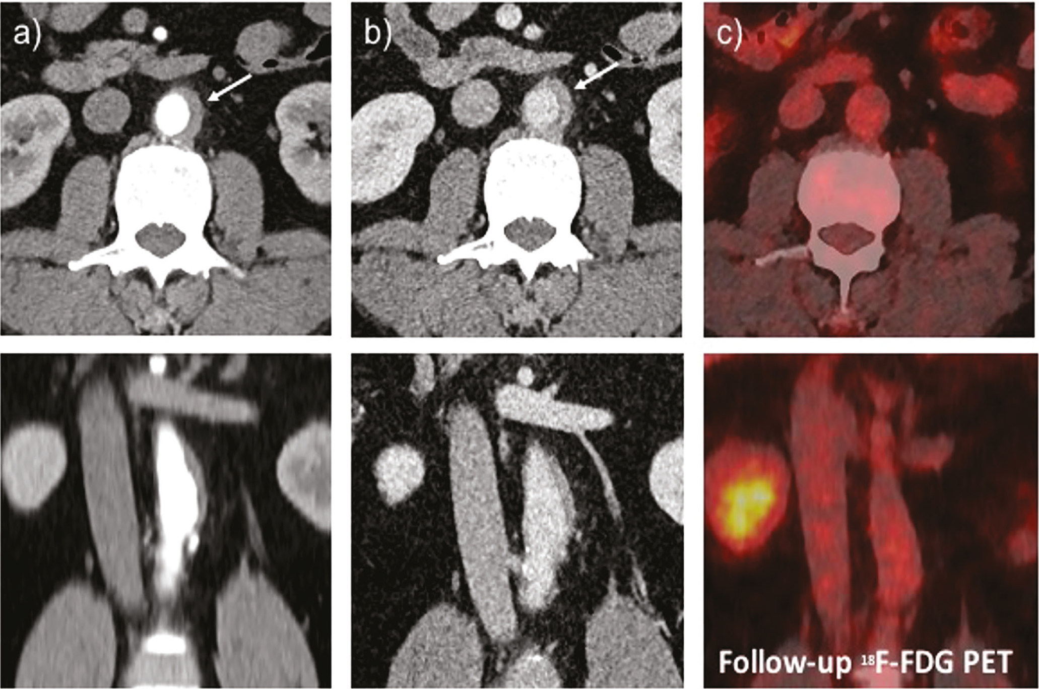
18F-fluorodeoxyglucose (18F-FDG) PET is widely utilized for diagnostic staging and restaging, assessing disease extent, monitoring treatment response, and supporting prognostic evaluation (,). 18F-FDG PET/CT serves not only as a tool for staging and treatment response assessment but also as a crucial modality for identifying clinically silent irAEs in patients undergoing chemoimmunotherapy (,). Recognizing these imaging patterns allows timely initiation of immunosuppressive therapy, helps prevent serious vascular complications, and supports the safer use of ICIs in oncology practice. This is particularly important in vascular irAEs, as untreated aortitis can progress to aneurysm formation, dissection, or even life-threatening rupture. Because clinical manifestations are often vague or absent, imaging becomes indispensable, and in this setting, 18F-FDG PET/CT assumes a key diagnostic role. 18F-FDG uptake corresponds to activated macrophages and lymphocytes within the inflamed aortic wall, allowing 18F-FDG PET/CT to identify active vasculitis even before structural changes develop. Several case series and imaging reviews have highlighted the role 18F-FDG PET/CT for detecting large-vessel inflammation in ICI-treated patients, outperforming contrast-enhanced CT when the inflammatory component is subtle and non-occlusive (,). This case highlights a rare but clinically significant presentation of nivolumab-induced periaortitis detected incidentally on 18F-FDG PET/CT during routine restaging. The characteristic linear aortic wall uptake prompted further evaluation with CT angiography, enabling early diagnosis and timely initiation of corticosteroid therapy. Complete metabolic resolution on follow-up PET/CT confirmed the immune-mediated nature of the process. As the use of ICIs continues to expand, awareness of vascular irAEs and the role of 18F-FDG PET/CT in their early detection is essential for preventing potentially serious complications.
Footnotes
Ethics
Informed consent: The consent was obtained for image use.
Authorship Contributions
Surgical and Medical Practices: S.U., S.P.N., Concept: S.U., N.A., Design: S.U., A.J., K.A.R., A.A.B., N.A., S.P.N., Data Collection or Processing: S.U., A.J., K.A.R., N.A., Analysis or Interpretation: S.U., K.A.R., S.P.N., Literature Search: S.U., Writing: S.U., S.P.N.
Conflict of Interest: No conflicts of interest were declared by the authors.
Financial Disclosure: The authors declare that this study has received no financial support.
References
Mellman I, Coukos G, Dranoff G. Cancer immunotherapy comes of age. Nature. 2011;480:480-489.
Chan KK, Bass AR. Autoimmune complications of immunotherapy: pathophysiology and management. BMJ. 2020;369:m736.
Usmani S, Ahmed DAT, Hamadah A, Al Kandari F. A rare case of diffuse large b-cell lymphoma of the prostate on 18F-FDG PET-CT. J Pak Med Assoc. 2021;71:388-389.
Prigent K, Aide N. 18F-fludeoxyglucose PET/computed tomography for assessing tumor response to immunotherapy and detecting immune-related side effects: a checklist for the PET reader. PET Clin. 2020;15:1-10.
Karlsen W, Akily L, Mierzejewska M, Teodorczyk J, Bandura A, Zaucha R, Cytawa W. Is 18F-FDG-PET/CT an optimal imaging modality for detecting immune-related adverse events after immune-checkpoint inhibitor therapy? Pros and Cons. Cancers (Basel). 2024;16:1990.
Usmani S, Jain A, Riyami KA, et al. Anti-programmed-death-receptor-1 (PD-1) antibody (pembrolizumab) induced pancreatitis diagnosed on 18F-FDG PET/CT. J Pak Med Assoc. 2024;74:825-826.
Hotta M, Naka G, Minamimoto R, Takeda Y, Hojo M. Nivolumab-induced periaortitis demonstrated by FDG PET/CT. Clin Nucl Med. 2020;45:910-912
Rossi G, Bauckneht M, Genova C, et al. Comparison between 18F-FDG PET-based and CT-based criteria in non-small cell lung cancer patients treated with nivolumab. J Nucl Med. 2020;61:990-998.

