

Abstract
Melorheostosis, a rare benign bone disorder marked by cortical thickening and irregular hyperostosis, typically affects peripheral bones but can also involve the axial skeleton. Though various molecular imaging radiotracers have been used to evaluate this condition, there are no previous reports of positive 68Ga-prostate-specific membrane antigen positron emission tomography/computed tomography (68Ga-PSMA PET/CT) findings in melorheostosis. This case presents a 70-year-old man with metastatic prostate cancer whose 68Ga-PSMA PET/CT scan revealed intense 68Ga-PSMA uptake in pubic melorheostosis seen on magnetic resonance imaging. The case highlights the possibility of 68Ga-PSMA-avid melorheostosis and suggests 68Ga-PSMA PET/CT may have a role in the evaluation of melorheostosis.
Keywords: Melorheostosis, 68Ga-PSMA, diagnostic challenge, PET/CT, prostate cancer, false positive
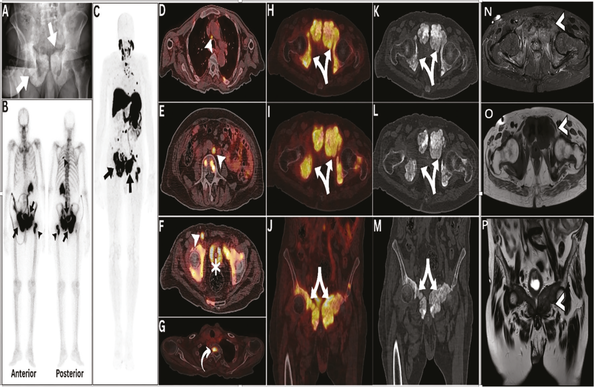
In our case; melorheostosis is located in the axial skeleton, more specifically in the pubic bones, which is a rare site of such entity (,). It also had increased 68Ga-PSMA activity, which can be misdiagnosed as bone metastases in patients with prostate cancer (). The observed expression pattern could be attributable to pronounced hypervascularization within melorheostotic lesions, consequently resulting in increased 68Ga-PSMA expression (,). Also, it can be attributable to the proposition that the RAS signaling pathway, which is implicated in melorheostosis, may interact with other pathways such as TGF-β and PI3K/AKT2 (). These pathways are known to regulate cellular processes like proliferation and differentiation, suggesting they could influence 68Ga-PSMA expression in this condition (). The differential diagnosis encompasses various osseous pathologies, including Paget’s disease, heterotopic ossification, and metastatic lesions (,,). Radiological findings showing a dripping candle wax appearance indicated melorheostosis in the patient (), highlighting the need for clinical follow-up and radiological correlation for accurate diagnosis. To the best of our knowledge, this case describes 68Ga-PSMA‑avid melorheostosis, reported here for the first time, and highlights its relevance as a pitfall in 68Ga-PSMA PET/CT interpretation, with the aim of preventing false upstaging of prostate cancer and avoiding unwarranted treatment.
Footnotes
Ethics
Informed Consent: The patient provided written informed consent.
Authorship Contributions
Surgical and Medical Practices: D.A.S., A.S.A., A.A-I., Concept: D.A.S., A.S.A., A.A-I., Design: D.A.S., A.S.A., A.A-I., Data Collection or Processing: D.A.S., A.S.A., A.A-I., Literature Search: D.A.S., A.S.A., A.A-I., Writing: D.A.S., A.S.A., A.A-I.
Conflict of Interest: No conflicts of interest were declared by the authors.
Financial Disclosure: The authors declare that this study has received no financial support.
References
Kalbermatten NT, Vock P, Rüfenacht D, Anderson SE. Progressive melorheostosis in the peripheral and axial skeleton with associated vascular malformations: imaging findings over three decades. Skeletal Radiol. 2001;30:48-52.
Suresh S, Muthukumar T, Saifuddin A. Classical and unusual imaging appearances of melorheostosis. Clin Radiol. 2010;65:593-600.
de Galiza Barbosa F, Queiroz MA, Nunes RF, Costa LB, Zaniboni EC, Marin JFG, Cerri GG, Buchpiguel CA. Nonprostatic diseases on PSMA PET imaging: a spectrum of benign and malignant findings. Cancer Imaging. 2020;20:23.
Fick CN, Fratzl-Zelman N, Roschger P, Klaushofer K, Jha S, Marini JC, Bhattacharyya T. Melorheostosis: a clinical, pathologic, and radiologic case series. Am J Surg Pathol. 2019;43:1554-1159.
Yao V, Berkman CE, Choi JK, O’Keefe DS, Bacich DJ. Expression of prostate-specific membrane antigen (PSMA), increases cell folate uptake and proliferation and suggests a novel role for PSMA in the uptake of the non-polyglutamated folate, folic acid. Prostate. 2010;70:305-316.
Sachpekidis C, Bäumer P, Kopka K, Hadaschik BA, Hohenfellner M, Kopp-Schneider A, Haberkorn U, Dimitrakopoulou-Strauss A. 68Ga-PSMA PET/CT in the evaluation of bone metastases in prostate cancer. Eur J Nucl Med Mol Imaging. 2018;45:904-912.
Jain TK, Vohra M, Sharma BS, Dhal S, Sharma LM. PSMA avidity in the heterotropic ossification—an incidental finding on PSMA PET/CT. Clin Nucl Med. 2024;49:e118-e9.
Sasikumar A, Joy A, Nanabala R, Pillai MRA, T.A H. 68Ga-PSMA PET/CT false-positive tracer uptake in paget disease. Clin Nucl Med. 2016;41:e454-e5.

