

Abstract
We report the case of a 31-year-old woman with osseous Hodgkin’s lymphoma who underwent post-therapeutic 18F-fluorodeoxyglucose (18F-FDG) positron emission tomography/computed tomography. The scan revealed a hypermetabolic lesion in the pubic symphysis and iliac crests, initially suggestive of residual disease. Multimodal evaluation, including 99mTc-methoxy-isobutyl-isonitrile scintigraphy, laboratory tests revealed primary hyperparathyroidism, and biopsy confirmed a brown tumor. This case highlights the importance of integrating multimodal imaging and pathology to avoid misinterpretation of 18F-FDG-avid lesions.
Keywords: Hodgkin’s lymphoma, brown tumor, primary hyperparathyroidism, 18F-FDG PET/CT, 99mTc-MIBI scintigraphy
,
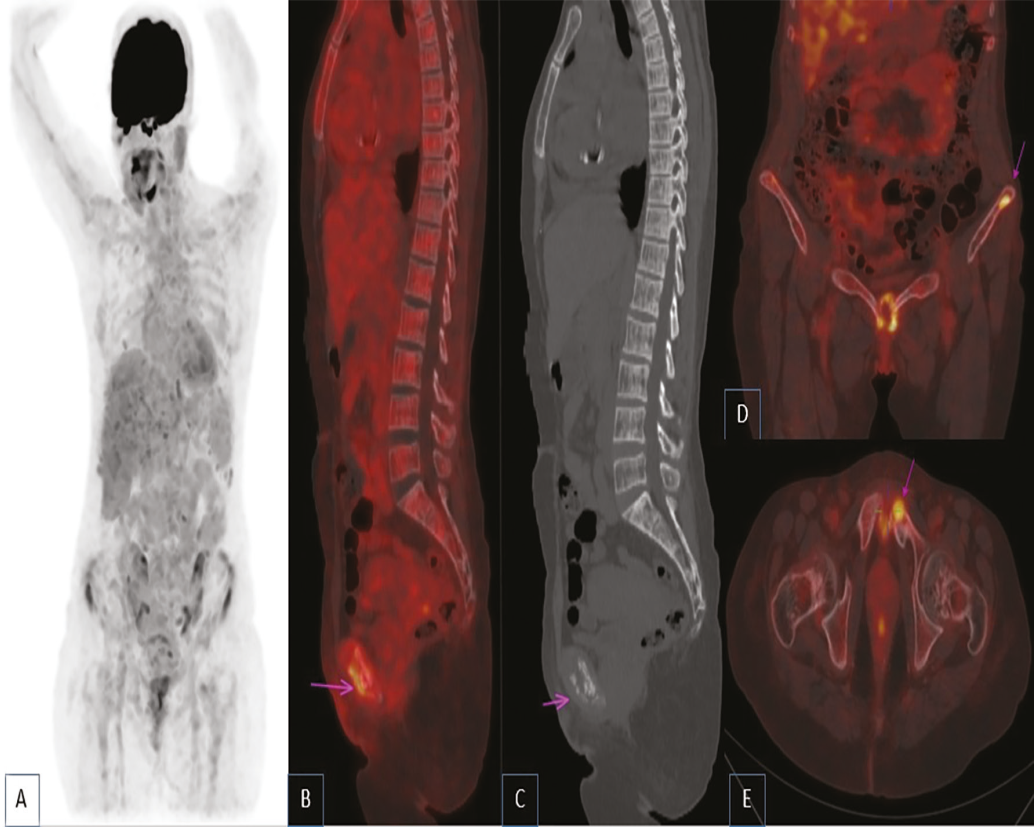
Further investigations were performed, as 18F-FDG-avid benign bone lesions, including brown tumors, can mimic residual malignancy (), particularly when the location is atypical for residual lymphoma.
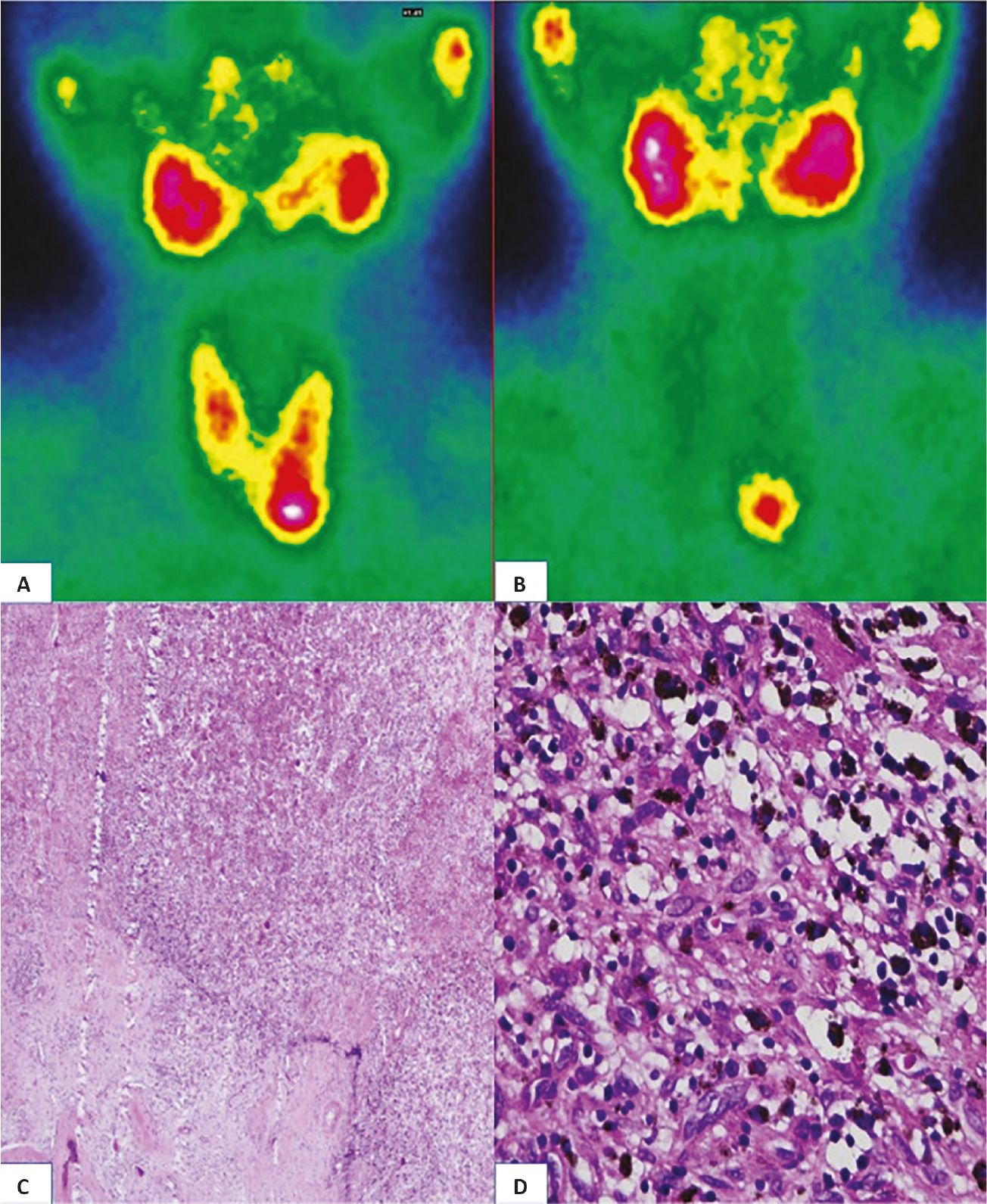
On 18F-FDG positron emission tomography/computed tomography, brown tumors may demonstrate intense metabolic activity and closely mimic malignant bone lesions. This increased 18F-FDG uptake reflects their high cellularity and increased osteoclastic activity, both of which are driven by excessive PTH stimulation. The resulting bone resorption leads to an accumulation of metabolically active fibroblasts and multinucleated giant cells with elevated glucose metabolism, explaining their strong 18F-FDG avidity. Similar diagnostic pitfalls have been reported, in which 18F-FDG-avid skeletal lesions were initially misinterpreted as metastases or giant-cell tumors before being correctly identified by biochemical assays and parathyroid scintigraphy (,). Although the coexistence of Hodgkin lymphoma and brown tumor is rare, previous reports have described benign 18F-FDG-avid bone lesions persisting after lymphoma remission, including giant cell tumor and fibrous dysplasia (,). Therefore, brown tumors and giant cell tumors should be considered in the differential diagnosis of 18F-FDG-avid bone lesions, particulary in the absence of biochemical or histopathological correlation.
Footnotes
Ethics
Informed Consent: The informed consent was obtained from the patient.
Authorship Contributions
Concept: D.N., A.M., S.M., I.G., H.G., Design: D.N., A.M., S.M., I.G., H.G., Data Collection or Processing: D.N., Analysis or Interpretation: D.N., A.M., H.G., Literature Search: D.N., Writing: D.N.
Conflict of Interest: No conflicts of interest were declared by the authors.
Financial Disclosure: The authors declare that this study has received no financial support.
References
Gladić Nenadić V, Punda M, Tabain A, Jukić T. Brown tumors in primary hyperparathyroidism initially interpreted as bone metastases and multiple gigantocelular tumors: case report. EJNMMI Rep. 2024;8:40.
Andersen KF, Albrecht-Beste E. Brown tumors due to primary hyperparathyroidism in a patient with parathyroid carcinoma mimicking skeletal metastases on (18)F-FDG PET/CT. Diagnostics (Basel). 2015;5:290-293.
Abdullahu LZ, Haxhiu BI, Orqusha NH, Gjonbalaj NA, Jashari AM, Kurshumliu FI, Fazliu VID, Lenjani BZ, Veseli SR, Kurtishi IN, Kaçiu YR. Brown tumor mimicking giant cell tumor in primary hyperparathyroidism: a case report. J Med Case Rep. 2025;19:429.
Dubreuil J, Salles G, Bozzetto J, Tordo J, Djaïleb L, Berriolo-Riedinger A, Leenhardt J, Giammarile F, Meignan M, Skanjeti A. Usual and unusual pitfalls of 18F-FDG-PET/CT in lymphoma after treatment: a pictorial review. Nucl Med Commun. 2017;38:563-576.
Gahier Penhoat M, Drui D, Ansquer C, Mirallie E, Maugars Y, Guillot P. Contribution of 18-FDG PET/CT to brown tumor detection in a patient with primary hyperparathyroidism. Joint Bone Spine. 2017;84:209-212

