

Abstract
Tumor-induced osteomalacia, resulting from tumoral overproduction of fibroblast growth factor 23, is a rare paraneoplastic syndrome clinically characterized by muscle weakness, bone pain, and pathological fractures. Fewer than 1,000 cases have been reported in the literature. Laboratory findings typically include hypophosphatemia, normal or decreased levels of 1.25-dihydroxy vitamin D, and normal or elevated levels of FGF23. Functional imaging methods, particularly somatostatin receptor imaging, play an important role in the localization of the tumor. In this case of tumor-induced osteomalacia, we presented the findings from gallium-68 (68Ga)-DOTATATE, 68Ga-fibroblast, and 18F-fluorodeoxyglucose positron emission tomography imaging modalities used for tumor localization.
Keywords: Tumor-induced osteomalacia, phosphaturic mesenchymal tumors, 68Ga-DOTATATE PET/CT, 68Ga-FAPI PET/CT, 18F-FDG PET/CT
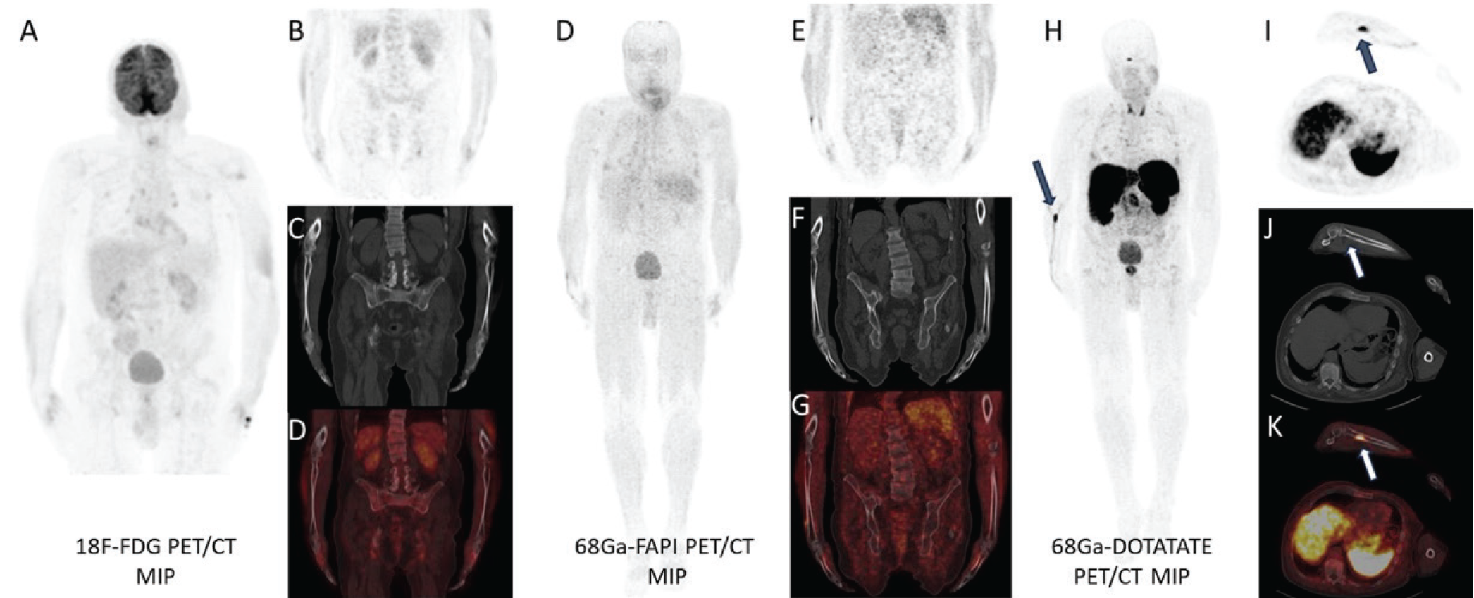
Tumor-induced osteomalacia (TIO) typically originates from phosphaturic mesenchymal tumors, which are usually small, slow-growing, and benign, and can be located in soft tissue or bone throughout the body from the skull to the feet (,). These tumors are most commonly localized in the lower extremities and the head and neck region, whereas the upper extremities are the least frequently affected sites (). Due to their typically small and solitary nature, identifying the exact location of these tumors is often challenging (). Localization generally begins with functional imaging techniques, including somatostatin receptor (SSTR) imaging (68Ga-DOTATATE or octreotide scan) and 18F-FDG PET/CT (). In a recent meta-analysis, SSTR-PET/CT demonstrated a higher detection rate for culprit tumors in TIO patients compared to 18F-FDG PET/CT (). Fibroblast activation protein inhibitor (FAPI) is known to accumulate in various benign and malignant conditions (,). Although only a limited number of TIO cases have been evaluated with FAPI imaging in the literature, some of these have shown uptake at the tumor site (). We evaluated the contribution of 68Ga-FAPI PET/CT, alongside 68Ga-DOTATATE PET/CT and 18F-FDG PET/CT, to tumor localization in a man with TIO. Our findings demonstrate the superiority of 68Ga-DOTATATE PET/CT over 18F-FDG and 68Ga-FAPI PET/CT for lesion localization.
Footnotes
Ethics
Informed Consent: Written informed consent was obtained from all patients before imaging.
Authorship Contributions
Concept: Z.T., U.E.S., İ.Ö., M.Y., B.E.A., Design: U.E.S., İ.Ö., M.Y., B.E.A., E.A., Data Collection or Processing: U.E.S., İ.Ö., M.Y., B.E.A., E.A., Analysis or Interpretation: U.E.S., İ.Ö., M.Y., B.E.A., E.A., Literature Search: U.E.S., İ.Ö., M.Y., B.E.A., E.A., Writing: Z.T., U.E.S., E.A.
Conflict of Interest: No conflicts of interest were declared by the authors.
Financial Disclosure: The authors declare that this study has received no financial support.
References
Florenzano P, Hartley IR, Jimenez M, et al. Tumor--induced osteomalacia. Calcif Tissue Int. 2021;108:128-142.
Hautmann AH, Hautmann MG, Kölbl O, et al. Tumor--induced osteomalacia: an up-to-date review. Curr Rheumatol Rep. 2015;17:512.
Bosman A, Palermo A, Vanderhulst J, et al. Tumor-induced osteomalacia: a systematic clinical review of 895 cases. Calcif Tissue Int. 2022;111:367-379.
Rayamajhi SJ, Yeh R, Wong T, et al. Tumor-induced osteomalacia - current imaging modalities and a systematic approach for tumor localization. Clin Imaging. 2019;56:114-123.
Jan de Beur SM, Minisola S, Xia WB, et al. Global guidance for the recognition, diagnosis, and management of tumor-induced osteomalacia. J Intern Med. 2023;293:309-328.
Meyer M, Nicod Lalonde M, Testart N, et al. Detection rate of culprit tumors causing osteomalacia using somatostatin receptor PET/CT: systematic review and meta-analysis. Diagnostics (Basel). 2019;10:2. Published 2019 Dec 18.
Tosunoğlu Z, Bozkurt M, Erdamar S, et al. Inflammatory 68Ga FAPI-46 uptake mimicking a sister Mary Joseph’s nodule in a case of gastric signet ring cell carcinoma. Clin Nucl Med. Published online May 14, 2025.
Şahin ÖF, Alçın G, Ergül N, Çermik TF, Arslan E. Physiological gallbladder accumulation on 68 Ga-FAPI-46 PET/CT. Clin Nucl Med. 2024;49:e685-e686.
Raeisi N, Saber Tanha A, Mosavi Z, et al. Phosphaturic mesenchymal tumor in the foot detected by 99mTc-FAPI-46 SPECT/CT and 18F-FDG PET/CT. Clin Nucl Med. Published online June 4, 2025.

