

Abstract
Dementia in individuals with Down syndrome (DS) is the leading cause of early-onset cognitive decline occurring before the age of 50. However, establishing an accurate diagnosis remains particularly challenging due to pre-existing intellectual disability, variability in baseline cognitive function, and the limited reliability of standard neuropsychological assessments. 18F-fluorodeoxyglucose positron emission tomography (18F-FDG PET) plays a critical role in identifying underlying neurodegenerative processes and supporting the diagnosis. We present a case of DS with multiple known and unknown pathologies identified on brain 18F-FDG PET and a confirmed diagnosis of early-onset mild Alzheimer’s disease.
Keywords: 18F-FDG, dementia, Alzheimer, Down syndrome
,
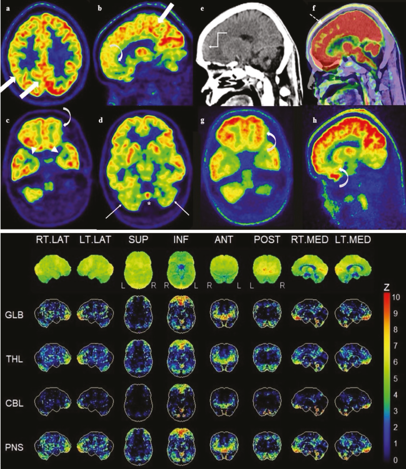
Markedly decreased uptake was detected bilaterally in the anteromedial temporal lobes, secondary to mild hippocampal atrophy (c; arrowheads).
Because of cerebellar vermian hypoplasia, uptake was decreased in the cerebellar hemispheres (d; thin arrows) and in the midline cerebellum (d; asterisk).
Decreased uptake was detected bilaterally in the anterior-inferior frontal lobes (b and c; right-curved arrow) without lobar atrophy on CT images (e; elbow-arrow). In the fused images, approximately 5 mm of head motion in the axial plane was detected between the CT and PET scans (f; dashed-arrow). Hypometabolism in the frontal lobes may have been an attenuation-correction artefact, but, in the light of non-attenuation-corrected (NAC) images, it is probably indicative of true hypometabolism (g and h; left-curved arrow).
The three-dimensional Stereotactic Surface Projections (3D-SSP) demonstrate the AD-related hypometabolic pattern by projecting stereotactically normalized 18F-FDG PET data onto a standardized cortical surface and displaying deviations from an age-matched normative database. Unfortunately, no biomarker test was performed.
DS dementia is the most common form of dementia under the age of 50-years (). Diagnosing dementia in Down syndrome (DS) could be challenging because of learning disabilities, unknown accuracy of cognitive tests, difficulties performing mental tests, and heterogeneity of intellectual functions.
Similar pattern of hypometabolism in AD diagnosed cases either sporadic or DS, suggesting similar neurodegeneration processes (). Although the diagnosis of AD was not confirmed with biomarker tests in our cases, a strong association between metabolic changes with biomarker levels was already reported (). 3D-SSP analysis which was proved to be useful in objectively revealing early metabolic changes confirmed AD (,,).
Neuroimaging presents some limitations in DS. Impaired communication may produce artifacts resulting from movement during acquisition. It has been reported that a spatial mismatch of 5-10 mm may result in errors of approximately 10-25% in measured 18F-FDG PET activity (,,). In our cases, we confirmed hypometabolism in the frontal lobes on NAC images. As a reflection difference in brain development, DS participants with or without dementia show lower metabolic rate and gray matter volume in frontal regions (). Besides, larger involvement of frontal regions in DS cases with AD than sporadic AD cases was also reported ().
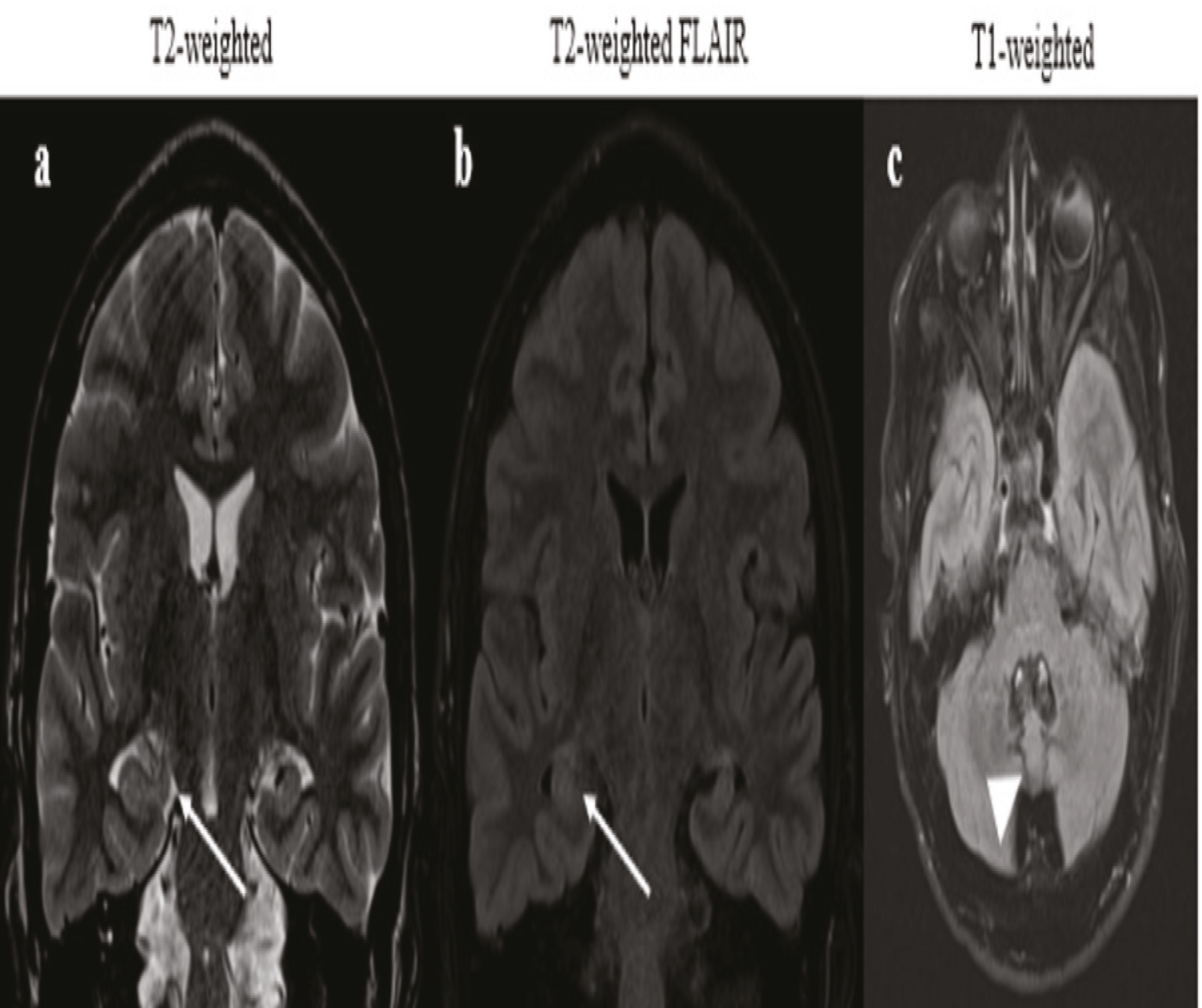
18F-FDG PET was critical not only because it revealed metabolic changes in anatomical pathologies that had already been detected by magnetic resonance imaging, but also because it clarified the AD diagnosis relevant to his current complaints. However, possible artefacts and variations must be well known in this specific patient group.
Footnotes
Ethics
Informed Consent: The written and verbal informed consent was obtained from the patient and his custodian.
Authorship Contributions
Surgical and Medical Practices: E.A., Concept: E.A., S.M., Design: E.A., S.M., Data Collection or Processing: E.A., Analysis or Interpretation: E.A., S.M., Literature Search: E.A., Writing: E.A.
Conflict of Interest: No conflicts of interest were declared by the authors.
Financial Disclosure: The authors declare that this study has received no financial support.
References
Hanney M, Prasher V, Williams N, Jones EL, Aarsland D, Corbett A, Lawrence D, Yu LM, Tyrer S, Francis PT, Johnson T, Bullock R, Ballard C; MEADOWS trial researchers. Memantine for dementia in adults older than 40 years with Down’s syndrome (MEADOWS): a randomised, double-blind, placebo-controlled trial. Lancet. 2012;379:528-536.
Arriola-Infante JE, Morcillo-Nieto AO, Zsadanyi SE, Franquesa-Mullerat M, Vaqué-Alcázar L, Rozalem-Aranha M, Arranz J, Rodríguez-Baz Í, Maure-Blesa L, Videla L, Barroeta I, Del Hoyo Soriano L, Benejam B, Fernández S, Sanjuan-Hernández A, Giménez S, Alcolea D, Belbin O, Flotats A, Camacho V, Lleó A, Carmona-Iragui M, Fortea J, Bejanin A. Regional brain metabolism across the Alzheimer’s disease continuum in Down syndrome. Ann Neurol. 2025;98:163-173.
Neale N, Padilla C, Fonseca LM, Holland T, Zaman S. Neuroimaging and other modalities to assess Alzheimer’s disease in Down syndrome. Neuroimage Clin. 2017;17:263-271.
Minoshima S, Frey KA, Koeppe RA, Foster NL, Kuhl DE. A diagnostic approach in Alzheimer’s disease using three-dimensional stereotactic surface projections of fluorine-18-FDG PET. J Nucl Med. 1995;36:1238-1248.
Burdette JH, Minoshima S, Vander Borght T, Tran DD, Kuhl DE. Alzheimer disease: improved visual interpretation of PET images by using three-dimensional stereotaxic surface projections. Radiology. 1996;198:837-843.
Andersson JL, Vagnhammar BE, Schneider H. Accurate attenuation correction despite movement during PET imaging. J Nucl Med. 1995;36:670-678.
Huang SC, Hoffman EJ, Phelps ME, Kuhl DE. Quantitation in positron emission computed tomography: 2. Effects of inaccurate attenuation correction. J Comput Assist Tomogr. 1979;3:804-814.
Kyme AZ, Fulton RR. Motion estimation and correction in SPECT, PET and CT. Phys Med Biol. 2021;66.
Sabbagh MN, Chen K, Rogers J, Fleisher AS, Liebsack C, Bandy D, Belden C, Protas H, Thiyyagura P, Liu X, Roontiva A, Luo J, Jacobson S, Malek-Ahmadi M, Powell J, Reiman EM. Florbetapir PET, FDG PET, and MRI in Down syndrome individuals with and without Alzheimer’s dementia. Alzheimers Dement. 2015;11:994-1004.

