

Abstract
Diffuse hepatic involvement in multiple myeloma is rare and may present a diagnostic challenge 18F-fluorodeoxyglucose positron emission tomography/computed tomography (18F-FDG PET/CT). We present the case of a 59-year-old man with a 3-year history of multiple myeloma who developed progressive hepatomegaly and perihepatic ascites. 18F-FDG PET/CT demonstrated marked diffuse liver enlargement without any focal or diffuse hepatic 18F-FDG uptake, while mild diffuse skeletal uptake suggested a globally 18F-FDG-low myeloma phenotype. Given the discordance between severe morphological findings and the absence of hepatic metabolic activity, a liver biopsy was performed, which revealed diffuse sinusoidal infiltration by CD138-positive, lambda-restricted plasma cells and excluded hepatic amyloidosis. Following systemic therapy, hepatomegaly and ascites regressed, while hepatic 18F-FDG uptake remained absent. Written informed consent for publication was obtained from the patient. This case highlights an important limitation of 18F-FDG PET/CT in detecting diffuse hepatic myeloma and emphasizes the need for multimodal diagnostic integration.
Keywords: 18F-FDG PET/CT, hepatic myeloma, diagnostic pitfall, multiple myeloma, hepatomegaly, liver biopsy
, ,
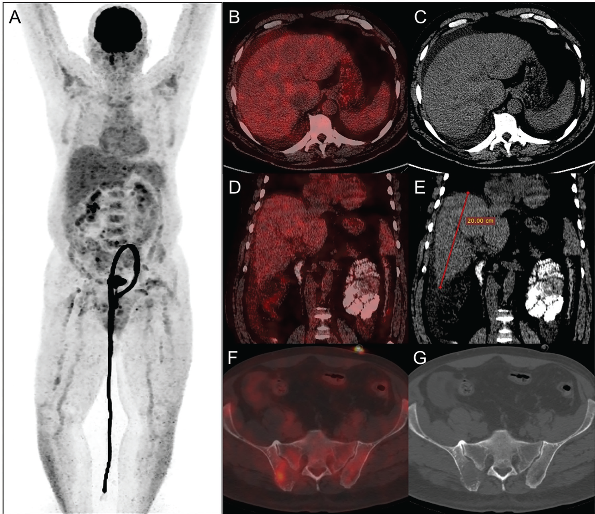
According to the IMWG-endorsed Italian Myeloma Criteria for PET Use (IMPeTUs), the imaging findings were consistent with a low-18F-FDG-avid myeloma phenotype. Bone marrow uptake demonstrated a Deauville score of 3 (BM3) with a mixed diffuse-focal pattern. Six lytic lesions were identified (L3 group), with the highest 18F-FDG uptake at the iliac bone biopsy site [maximum standardized uptake value (SUVmax): 4.8], comparable to hepatic reference activity (SUVmax: 4.1). No paramedullary or extramedullary metabolically active disease was detected. Overall, the IMPeTUs profile was classified as BM3 Sp F2 L3. Bone marrow biopsy demonstrated marked plasma-cell infiltration (approximately 85% of marrow cellularity) with CD138 and MUM1 positivity and lambda light-chain restriction (kappa/lambda ratio 1/80), consistent with relapsed plasma cell myeloma.
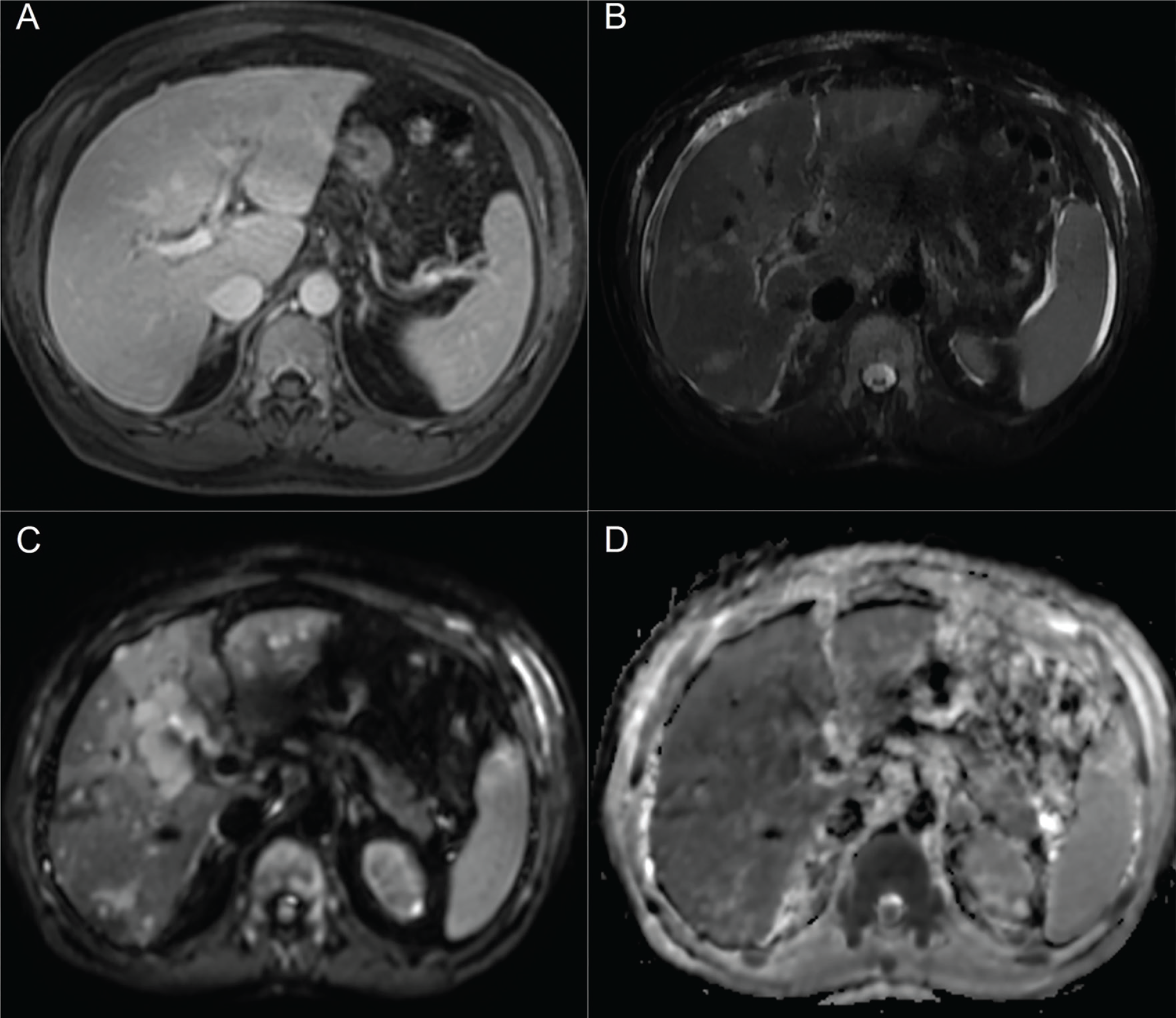
Hepatic involvement in multiple myeloma is uncommon, and tumor-forming hepatic lesions are particularly rare. Reported histopathological patterns include extramedullary plasmacytoma, light-chain deposition disease, amyloidosis, and diffuse infiltrative forms such as sinusoidal plasma-cell infiltration, which may present radiologically as hepatomegaly with ascites in the absence of discrete focal lesions ().
The striking discrepancy between severe hepatomegaly with ascites and complete absence of hepatic 18F-fluorodeoxyglucose uptake prompted further diagnostic evaluation. Percutaneous liver biopsy demonstrated diffuse sinusoidal infiltration by plasma cells with strong CD138 expression and lambda light-chain restriction, confirming hepatic involvement by multiple myeloma. Congo red staining was negative, excluding hepatic amyloidosis (,).
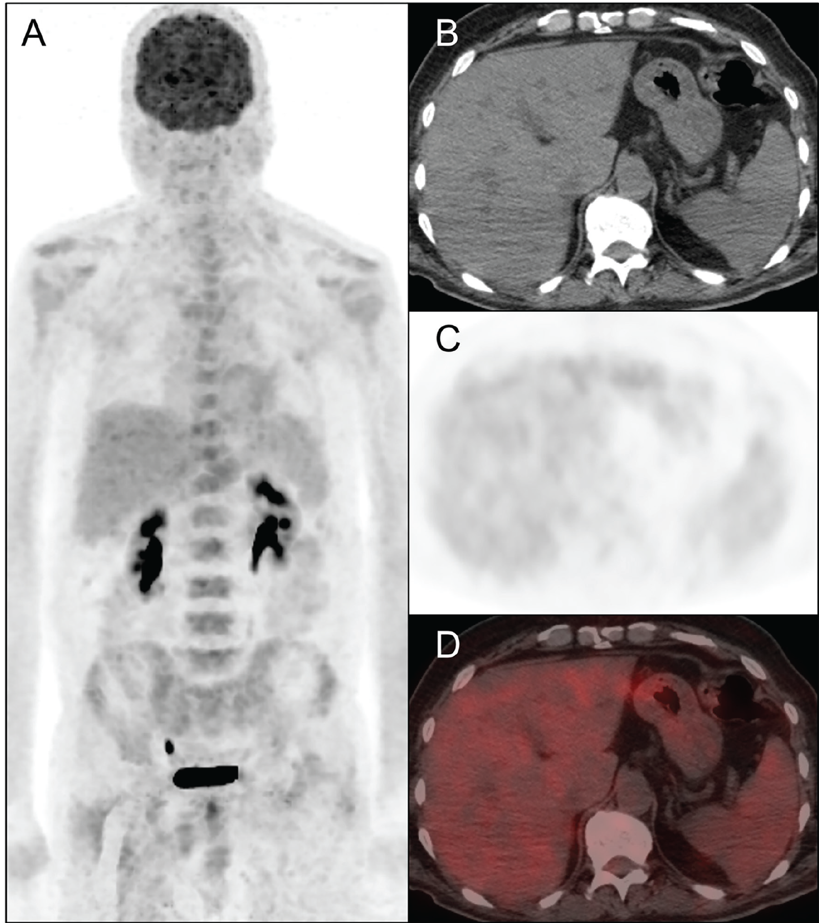
Hepatic involvement in multiple myeloma is uncommon, with reported frequencies ranging from approximately 0.3-3%, and diffuse infiltrative patterns are far less frequently encountered than focal plasmacytomas. Histopathological manifestations include diffuse sinusoidal plasma-cell infiltration, light-chain deposition disease, amyloidosis, and less commonly tumor-forming lesions. This case illustrates a well-recognized but underappreciated limitation of 18F-FDG PET/CT in detecting diffuse hepatic involvement; low 18F-FDG avidity in such presentations has been proposed to relate to reduced hexokinase-2 expression and alternative metabolic pathways in certain plasma-cell phenotypes, as supported by prior translational and imaging studies rather than being directly demonstrated in the present case (,,). In patients presenting with unexplained hepatomegaly and discordant metabolic imaging findings, histopathological confirmation remains essential. In this context, alternative PET tracers most notably CXCR4-targeted agents and, more recently, fluorocholine PET may provide complementary information in 18F-FDG -low myeloma phenotypes (,,).
Footnotes
Ethics
Informed Consent: Written informed consent for publication was obtained from the patient.
Authorship Contributions
Surgical and Medical Practices: S.B., Concept: S.B., E.A.İ., Design: S.B., M.O., E.A.İ., Data Collection or Processing: S.B., E.A.İ., Analysis or Interpretation: S.B., E.A.İ., Literature Search: M.O., E.A.İ., Writing: S.B., M.O., E.A.İ.
Conflict of Interest: No conflicts of interest were declared by the authors.
Financial Disclosure: The authors declare that this study has received no financial support.
References
Cavo M, Terpos E, Nanni C, Moreau P, Lentzsch S, Zweegman S, Hillengass J, Engelhardt M, Usmani SZ, Vesole DH, San-Miguel J, Kumar SK, Richardson PG, Mikhael JR, da Costa FL, Dimopoulos MA, Zingaretti C, Abildgaard N, Goldschmidt H, Orlowski RZ, Chng WJ, Einsele H, Lonial S, Barlogie B, Anderson KC, Rajkumar SV, Durie BGM, Zamagni E. Role of 18 F-FDG PET/CT in the diagnosis and management of multiple myeloma and other plasma cell disorders: a consensus statement by the International Myeloma Working Group. Lancet Oncol. 2017;18:e206-e217.
Tirumani SH, Sakellis C, Jacene H, Shinagare AB, Munshi NC, Ramaiya NH, Van den Abbeele AD. Role of FDG-PET/CT in extramedullary multiple myeloma: correlation of FDG-PET/CT findings with clinical outcome. Clin Nucl Med. 2016;41:e7-e13.
Tirotta D, Lena C, Torre M, Muratori P. Hematologic focal hepatic lesions in the absence of liver cirrhosis: consider lymphoma and myeloma. Clin Res Hepatol Gastroenterol. 2026;50:102749.
Tomasian A, Sandrasegaran K, Elsayes KM, Shanbhogue A, Shaaban A, Menias CO. Hematologic malignancies of the liver: spectrum of disease. Radiographics. 2015;35:71-86.
Pu Z, Zhan L, Zha Y, Li Y, Qiu S, He Y, Yang Y, Feng Z, Zhu X. Hepatic involvement as an initial manifestation of multiple myeloma: a case report with atypical imaging features. Front Oncol. 2025;15:1640981.
Singh M, Singh H, Pham P, Rizvi B, Rao R. Extramedullary multiple myeloma with hepatic involvement. Cureus. 2021;13:e13515.
Abe Y, Ikeda S, Kitadate A, Narita K, Kobayashi H, Miura D, Takeuchi M, O’uchi E, O’uchi T, Matsue K. Low hexokinase-2 expression-associated false-negative 18 F-FDG PET/CT as a potential prognostic predictor in patients with multiple myeloma. Eur J Nucl Med Mol Imaging. 2019;46:1345-1350.
Rasche L, Angtuaco E, McDonald JE, Buros A, Stein C, Pawlyn C, Thanendrarajan S, Schinke C, Samant R, Yaccoby S, Walker BA, Epstein J, Zangari M, van Rhee F, Meissner T, Goldschmidt H, Hemminki K, Houlston R, Barlogie B, Davies FE, Morgan GJ, Weinhold N. Low expression of hexokinase-2 is associated with false-negative FDG-positron emission tomography in multiple myeloma. Blood. 2017;130:30-34.
Zamagni E, Patriarca F, Nanni C, Zannetti B, Englaro E, Pezzi A, Tacchetti P, Buttignol S, Perrone G, Brioli A, Pantani L, Terragna C, Carobolante F, Baccarani M, Fanin R, Fanti S, Cavo M. Prognostic relevance of 18-F FDG PET/CT in newly diagnosed multiple myeloma patients treated with up-front autologous transplantation. Blood. 2011;118:5989-5995.
Shekhawat AS, Singh B, Malhotra P, Watts A, Basher R, Kaur H, Hooda M, Radotra BD. Imaging CXCR4 receptors expression for staging multiple myeloma by using 68 Ga-pentixafor PET/CT: comparison with 18 F-FDG PET/CT. Br J Radiol. 2022;95:20211272.
Kaya G, Akin S, Buyukasik Y, Bozkurt MF, Tuncel M, Kiratli PO. PET in myeloma redefined: a comparative imaging study with FDG and fluorocholine PET/CT. Eur J Nucl Med Mol Imaging. 2026;53:2325-2336.
Chavoshi M, Mirshahvalad SA, Kohan A, Ortega C, Metser U, Farag A, Kridel R, Hodgson D, Bhella S, Kukreti V, Veit-Haibach P. CXCR4-targeted PET imaging in hematologic malignancies: a systematic review and meta-analysis. Clin Nucl Med. 2025;50:e7-e16.

