
68Ga-PSMA PET/CT Detects a Rare Case of Breast Metastasis from Prostate Cancer
PDF

Interesting Image

68Ga-PSMA PET/CT Detects a Rare Case of Breast Metastasis from Prostate Cancer

Mol Imaging Radionucl Ther 2026;35(2):120-122 DOI: 10.4274/mirt.galenos.2025.98475
, , , ,
PDF
Permissions

Abstract

Positron emission tomography/computed tomography (PET/CT) with Galium-68 prostate-specific membrane antigen (68Ga-PSMA) PET/CT) is a valuable tool for initial staging, recurrence detection, and assessment of treatment response in men with prostate cancer. Metastatic spread to regional and distant lymph nodes, bone, lung and liver is well known and expected in patients with castration-resistant disease. However, the application of a highly sensitive whole-body imaging modality such as 68Ga-PSMA PET/CT can reveal metastatic foci in rare or unusual locations. We present the case of an 82-year-old patient with prostate adenocarcinoma who underwent long-term androgen deprivation therapy. A 68Ga-PSMA PET/CT scan was performed in June 2021 due to significant prostate-specific antigen elevation and revealed disease progression with widespread regional and distant metastases, including a soft-tissue mass in the right breast that demonstrated intense radiotracer uptake; this mass was subsequently resected and histologically confirmed. This case underscores the excellent diagnostic performance of 68Ga-PSMA PET/CT for atypical metastatic sites.

Keywords: Prostate cancer, breast metastasis, 68Ga-PSMA PET/CT
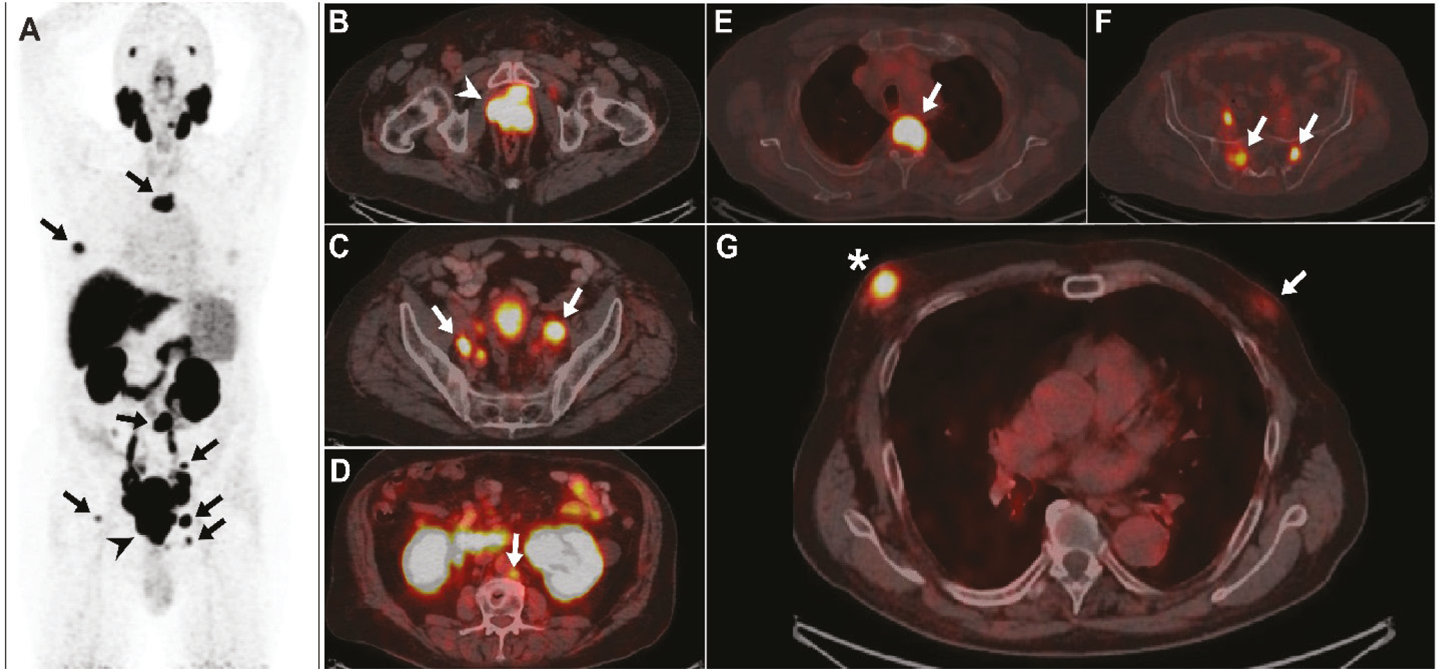
Figure 1
Figure 1. An 82-year-old patient, who had been diagnosed with high-grade prostate adenocarcinoma in November 2014 (then aged 75) with a Gleason score of 8 (4+4) and a pre-biopsy serum prostate-specific antigen (PSA) concentration of 135 µg/L, was admitted to our department for further evaluation. Initial staging with bone scintigraphy and abdominopelvic computed tomography (CT) revealed locally advanced disease, without evidence of regional or distant dissemination. The patient underwent long-term androgen deprivation therapy until June 2021, when he was referred for restaging with Galium-68 prostate-specific membrane antigen (68Ga-PSMA) positron emission tomography (PET)/CT because of a significant PSA elevation (182 µg/L). Prior to the scan, the patient reported a painful right breast lump that he had noticed one month earlier. The scan was performed after obtaining a written consent and in accordance with the standard protocol, 60 min after intravenous administration of 4.5 mCi 68Ga-PSMA-11 (). Maximum-intensity projection images demonstrated intense PSMA uptake in the prostate gland (A, arrowhead) and in multiple metastatic sites (A, arrows). Fused images revealed an enlarged prostate with lobulated margins and intense PSMA uptake throughout the gland (B, arrowhead). Multiple PSMA-avid parailiac and paraaortic lymph nodes (C and D; arrows) and several bone lesions (E and F; arrows) were identified. Remarkably, there was evidence of a focal soft-tissue lesion in the right breast with intense uptake, measuring 23 × 20 mm in axial diameter (G, asterisk). Increased subareolar glandular tissue with faint activity was also noted in the contralateral left breast (G; arrow). Following the PET/CT, the patient was referred for further evaluation with mammography, which demonstrated benign gynecomastia in the left breast and a lesion with malignant characteristics in the right breast. To definitively exclude a primary breast tumor, the patient was referred for surgical excision of the lesion. Histopathological examination and immunohistochemistry confirmed the presence of breast metastasis from prostate carcinoma. Breast metastases from prostate cancer are uncommon, with only a few cases reported in the literature (). Long-term hormonal treatment is considered a predisposing factor, often causing gynecomastia and increased breast vascularity, which facilitate hematogenous metastatic spread (). Kumar Chauhan et al. () report a similar case of a patient with metastatic castration-resistant prostate cancer (after surgical castration and anti-androgen therapy) who had a metastasis in the left breast that was also PSMA-positive, considerably larger, and even infiltrated the adjacent chest wall. Previous studies have shown that increased PSMA expression can also occur in benign changes associated with gynecomastia, which may be unilateral or asymmetric (,). Synchronous breast cancer should also be considered, given the increased expression of PSMA in the tumor-associated neovasculature of various non-prostatic malignancies. Polverari et al. () described a PSMA-avid breast lesion detected on restaging PET/CT that was confirmed histologically as primary male breast cancer. In contrast, their patient presented with an isolated locoregional recurrence after radical prostatectomy (a PSMA-positive para-iliac lymph node) rather than widespread metastatic prostate cancer and had no prior anti-androgen therapy. Our report underscores the excellent diagnostic performance of 68Ga-PSMA PET/CT for the detection of unusual metastatic sites and emphasizes potential pitfalls in the interpretation of such findings.
View larger Download

Footnotes

Ethics

Informed Consent: Informed consent was obtained from the patient for the publication of his anonymized images and relevant clinical information.

Authorship Contributions

Concept: G.G., Design: N.H., V.H., Data Collection or Processing: M.I., N.H.,  Analysis or Interpretation: P.N., M.I., V.H.,  Literature Search: P.N., M.I., Writing: G.G.

Conflict of Interest: No conflicts of interest were declared by the authors.

Financial Disclosure: The authors declare that this study has received no financial support.

References

1

Fendler WP, Eiber M, Beheshti M, Bomanji J, Calais J, Ceci F, Cho SY, Fanti S, Giesel FL, Goffin K, Haberkorn U, Jacene H, Koo PJ, Kopka K, Krause BJ, Lindenberg L, Marcus C, Mottaghy FM, Oprea-Lager DE, Osborne JR, Piert M, Rowe SP, Schöder H, Wan S, Wester HJ, Hope TA, Herrmann K. PSMA PET/CT: joint EANM procedure guideline/SNMMI procedure standard for prostate cancer imaging 2.0. Eur J Nucl Med Mol Imaging. 2023;50:1466-1486.

2

Zhang S, Peng W, Cheng L, Pei B, Zhou X, Wang M, Jiang L, Lu C, Xu L. Metastasis of prostate cancer to breast: a case report. Front Oncol. 2025;15:1580441.

3

Lima V, Martinez-Lapus FG, Demegillo KJ. Breast metastasis from castrate-resistant prostatic adenocarcinoma mimicking as a second primary: a case report. World J Oncol. 2020;11:37-40.

4

Kumar Chauhan P, Jain M, Adiga P, Shivalingaiah M. Breast lump: an uncommon metastasis of prostate cancer. Archives of Clinical and Medical Case Reports. 2020;4:624-628.

5

Sasikumar A, Joy A, Nair BP, Pillai MRA, Madhavan J. False positive uptake in bilateral gynecomastia on 68Ga-PSMA PET/CT scan. Clin Nucl Med. 2017;42:e412-e414.

6

Metwalley KA, Farghaly HS. Gynecomastia in adolescent males: current understanding of its etiology, pathophysiology, diagnosis, and treatment. Ann Pediatr Endocrinol Metab. 2024;29:75-81.

7

Polverari G, Ceci F, Calderoni L, Cervati V, Farolfi A, Castellucci P, Fanti S. Male breast cancer detected by 68 Ga-PSMA-11 PET/CT in a patient with prostate cancer with pelvic lymph node metastasis. Clin Genitourin Cancer. 2019;17:154-156.

