

Abstract
We present a case of a 40-year-old male with refractory multiple myeloma, assessed using 68Gallium-PENTIXAFOR positron emission tomography/computed tomography (68Ga-PENTIXAFOR PET/CT), revealing widespread and intense C-X-C motif chemokine receptor 4 (CXCR4) expression in multiple osteolytic lesions across axial and appendicular skeletons, including bone marrow deposits. Despite undergoing autologous hematopoietic stem cell transplantation and multiple lines of maintenance therapy, the patient experienced disease relapse and progression. The term “superscan” typically refers to diffuse skeletal uptake in conventional bone scans, primarily seen in advanced metastatic cancers or metabolic bone diseases. CXCR4, crucial for tumor growth and metastasis, binds C-X-C motif chemokine 12 (CXCL12) to promote cancer progression. PENTIXAFOR, a CXCR4-targeted PET agent, facilitates imaging of such malignancies. While superscans using PET/CT are rare, our case underscores the utility of 68Ga-PENTIXAFOR PET/CT in evaluating CXCR4 expression in multiple myeloma, highlighting its potential as a diagnostic and prognostic tool in refractory disease management.
Keywords: PENTIXAFOR, C-X-C motif chemokine receptor 4, positron emission tomography/computed tomography, superscan, multiple myeloma
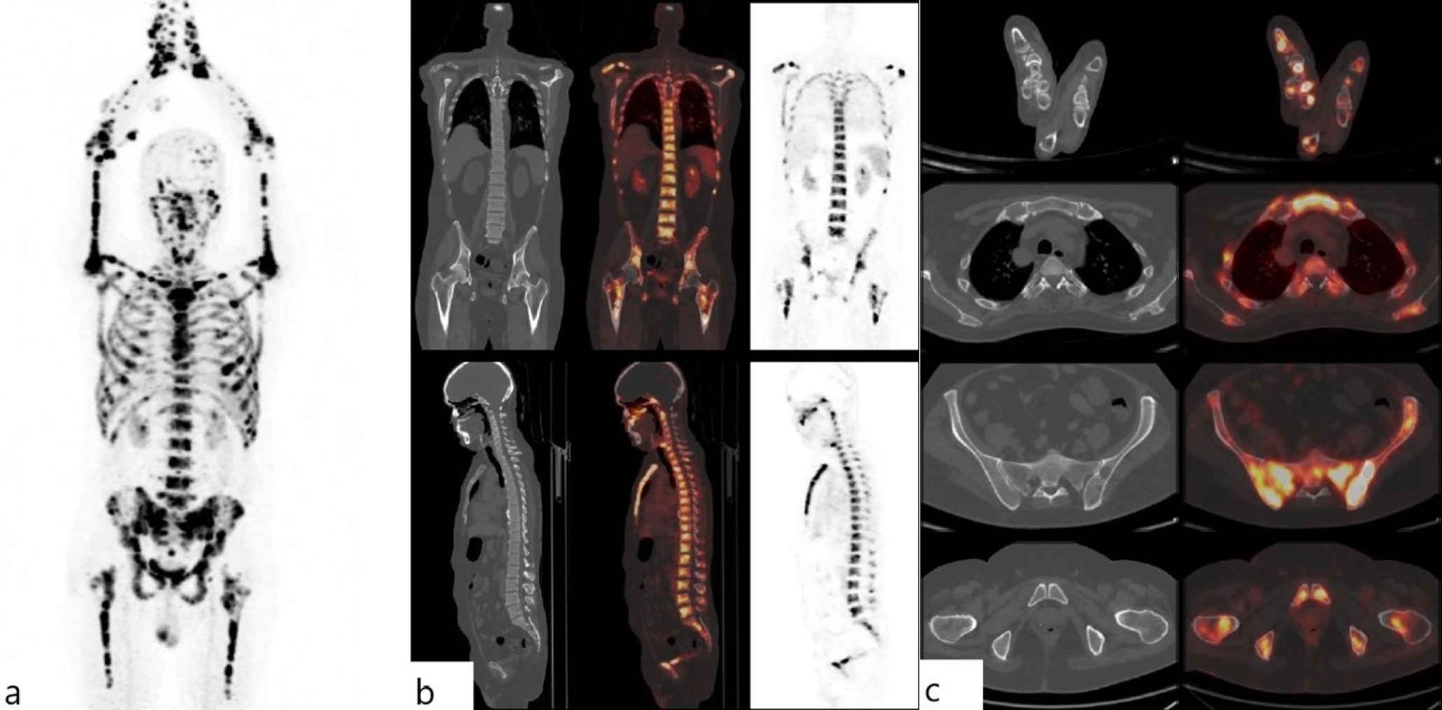
Footnotes
Ethics
Informed Consent: Patient consent was obtained for this study.
Authorship Contributions
Concept: V.R.L., Design: S.K., H.G., Data Collection or Processing: S.K., N.S., Analysis or Interpretation: N.S., Literature Search: S.K., H.G., Writing: S.K.
Conflict of Interest: No conflicts of interest were declared by the authors.
Financial Disclosure: The authors declare that this study has received no financial support.
References
Jacobson O, Weiss ID. CXCR4 chemokine receptor overview: biology, pathology and applications in imaging and therapy. Theranostics. 2013; 3:1-2.
Chen J, Xu-Monette ZY, Deng L, Shen Q, Manyam GC, Martinez-Lopez A, Zhang L, Montes-Moreno S, Visco C, Tzankov A, Yin L, Dybkaer K, Chiu A, Orazi A, Zu Y, Bhagat G, Richards KL, Hsi ED, Choi WW, van Krieken JH, Huh J, Ponzoni M, Ferreri AJ, Zhao X, Møller MB, Farnen JP, Winter JN, Piris MA, Pham L, Young KH. Dysregulated CXCR4 expression promotes lymphoma cell survival and independently predicts disease progression in germinal center B-cell-like diffuse large B-cell lymphoma. Oncotarget. 2015;6:5597-614.
Shekhawat AS, Singh B, Malhotra P, Watts A, Basher R, Kaur H, Hooda M, Radotra BD. Imaging CXCR4 receptors expression for staging multiple myeloma by using 68Ga-Pentixafor PET/CT: comparison with 18F-FDG PET/CT. Br J Radiol. 2022;95:20211272.
Kraus S, Dierks A, Rasche L, Kertels O, Kircher M, Schirbel A, Zovko J, Steinbrunn T, Tibes R, Wester HJ, Buck AK, Einsele H, Kortüm KM, Rosenwald A, Lapa C.Ga-Pentixafor PET/CT for detection of chemokine receptor CXCR4 expression in myeloproliferative neoplasms. J Nucl Med. 2022; 63:96-99.
Hu J, Xu T, Shen H, Song Y, Yang J, Zhang A, Ding H, Xing N, Li Z, Qiu L, Ma L, Yang Y, Feng Z, Du Z, He W, Sun Y, Cai J, Li Q, Chen Y, Yang S. Chongqing Primary Aldosteronism Study (CONPASS) Group. Accuracy of Gallium-68 pentixafor positron emission tomography-computed tomography for subtyping diagnosis of primary aldosteronism. JAMA Netw Open. 2023; 6(2):e2255609.
Chen Z, Yang A, Chen A, Dong J, Lin J, Huang C, Zhang J, Liu H, Zeng Z, Miao W. [Ga]Pentixafor PET/CT for staging and prognostic assessment of newly diagnosed multiple myeloma: comparison to [F]FDG PET/CT. Eur J Nucl Med Mol Imaging. 2024; 51:1926-1936.
Fu Z, Chen X, Yang X, Li Q. Skeletal Superscan on 18F-FDG PET/CT in a patient with multiple myeloma. Clin Nucl Med. 2019; 44:169-170.
Pan Q, Luo Y, Cao X, Ma Y, Li F. Multiple myeloma presenting as a superscan on 68ga-pentixafor PET/CT. Clin Nucl Med. 2018; 43:462-463.

