

Abstract
Fibroblast activation protein (FAPI), a type II transmembrane glycoprotein is a promising target to image epithelial originated cancers. Pancreatic cancer is characterized with [68Ga]Ga-FAPI-04 and 18F-fluorodeoxyglucose(18F-FDG) uptake in varying degree. However physiologic uptake and uptake associated with acute/chronic pancreatitis makes interpretation challenging. We would like to present a case of pancreatic cancer whose tumor could not delineated from rest pancreatic tissue in [68Ga]Ga-FAPI-04 positron emission tomography/computed tomography (PET/CT) images due to intense FAPI uptake in whole pancreas but more remarkable in 18F-FDG PET/CT images.
Keywords: FAPI, Gallium-68, pancreatic cancer, FDG, PET/CT
,
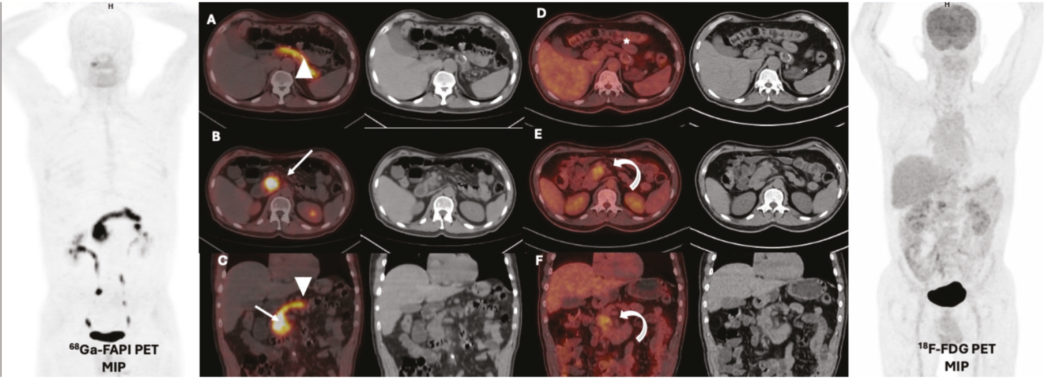
In this case due to high [68Ga]Ga- fibroblast activation protein (FAPI)-04 uptake in the whole pancreatic tissue delineating the tumor from non-cancerous parenchyma was not possible. Although tumor showed low degree hypermetabolism, the tumor border was clearer in 18F-fluorodeoxyglucose positron emission tomography (18F-FDG PET) images compared with [68Ga]Ga-FAPI-04 PET images.
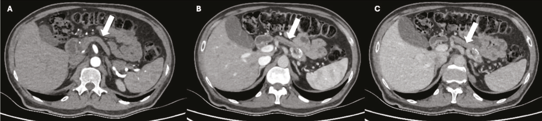
[68Ga]Ga-FAPI-04 has several advantages over 18F-FDG PET in terms of patient preparation. [68Ga]Ga-FAPI-04 PET is a promising alternative to 18F-FDG PET in cases with uncontrolled hyperglycemia like ours’. Superiority of [68Ga]Ga-FAPI-04 PET in most epithelial cancer originating from the gastrointestinal tract especially in identifying lymph nodes and peritoneal metastasis have shown in the literature (-). Similar to 18F-FDG, both focal and diffuse [68Ga]Ga-FAPI-04 uptakes are a pitfall in cases with pancreatic carcinoma suspicion. Non-cancerous pathology of the pancreas could show high [68Ga]Ga-FAPI-04 uptake (,). Moreover, tumor induced acute pancreatitis made more complex the delineation of the tumor (). Non-specific prominent [68Ga]Ga-FAPI-04 uptake compared with 18F-FDG could be detected probably due to fibrotic or chronic inflammatory changes of pancreas (). In most cases, the physiologic [68Ga]Ga-FAPI-04 uptake of pancreas is lower compared with 18F-FDG (). There is limited data in the literature suggesting that high-degree [68Ga]Ga-FAPI-04 uptake in pancreatitis may mask the tumor (). Our case was diagnosed with diabetes and pancreas appeared atrophic on CT images. Both findings could support chronic pancreatitis. In our case, diffuse intense [68Ga]Ga-FAPI-04 uptake and low-grade 18F-FDG uptake were thought to be secondary to chronic pancreatitis inducing fibrosis. [68Ga]Ga-FAPI-04 uptake in the fibrotic tissue is well known in literature. In selected cases, dual-tracer PET imaging could increase our knowledge about the cancer and concomitant pathologies.
Footnotes
Ethics
Informed Consent: Informed consent was obtained from all subjects involved in the study.
Authorship Contributions
Surgical and Medical Practices: E.A., A.E.Ö., G.A., M.M.S., Es.A., Concept: E.A., Design: E.A., Es.A., Data Collection or Processing: E.A., M.M.S., Analysis or Interpretation: E.A., M.M.S., Literature Search: E.A., A.E.Ö., Writing: E.A.
Conflict of Interest: No conflicts of interest were declared by the authors.
Financial Disclosure: The authors declare that this study has received no financial support.
References
Giesel FL, Kratochwil C, Schlittenhardt J, Dendl K, Eiber M, Staudinger F, Kessler L, Fendler WP, Lindner T, Koerber SA, Cardinale J, Sennung D, Roehrich M, Debus J, Sathekge M, Haberkorn U, Calais J, Serfling S, Buck AL. Head-to-head intra-individual comparison of biodistribution and tumor uptake of 68Ga-FAPI and 18F-FDG PET/CT in cancer patients. Eur J Nucl Med Mol Imaging. 2021;48:4377-4385.
Pang Y, Zhao L, Shang Q, Meng T, Zhao L, Feng L, Wang S, Guo P, Wu X, Lin Q, Wu H, Huang W, Sun L, Chen H. Positron emission tomography and computed tomography with [68Ga]Ga-fibroblast activation protein inhibitors improves tumor detection and staging in patients with pancreatic cancer. Eur J Nucl Med Mol Imaging. 2021;49:1322-1337.
Pang Y, Zhao L, Luo Z, Hao B, Wu H, Lin Q, Sun L, Chen H. Comparison of 68Ga-FAPI and 18F-FDG uptake in gastric, duodenal, and colorectal cancers. Radiology. 2021;298:393-402.
Gündoğan C, Kömek H, Can C, Yildirim ÖA, Kaplan İ, Erdur E, Poyraz K, Güzel Y, Oruç Z, Çakabay B. Comparison of 18F-FDG PET/CT and 68Ga-FAPI-04 PET/CT in the staging and restaging of gastric adenocarcinoma. Nucl Med Commun. 2022;43:64-72.
Veldhuijzen van Zanten SEM, Pieterman KJ, Wijnhoven BPL, Pruis IJ, Groot Koerkamp B, van Driel LMJW, Verburg FA, Thomeer MGJ. FAPI PET versus FDG PET, CT or MRI for staging pancreatic-, gastric- and cholangiocarcinoma: systematic review and head-to-head comparisons of diagnostic performances. Diagnostics (Basel). 2022;12:1958.
Shou Y, Xue Q, Yuan J, Zhao J. 68Ga-FAPI-04 PET/MR is helpful in differential diagnosis of pancreatitis from pancreatic malignancy compared to 18F-FDG PET/CT: a case report. Eur J Hybrid Imaging. 2021;5:12.
Luo Y, Pan Q, Zhang W, Li F. Intense FAPI uptake in ınflammation may mask the tumor activity of pancreatic cancer in 68Ga-FAPI PET/CT. Clin Nucl Med. 2020;45:310-311.
Li Y, Gao J, Li Y, Duan X, Shen C. Non-specific uptake of 18F-FAPI-04 in the pancreas and its related factors: a post-hoc analysis of an ongoing prospective clinical trial. Sci Rep. 2024;14:11141.

