

Abstract
Spondyloepiphyseal dysplasia tarda (SEDT) is a rare X-linked skeletal disorder affecting the spine and long bones, leading to short stature, spinal deformities, and joint stiffness. It is caused by genetic mutations, and primarily affects males. Diagnosis is confirmed by imaging and genetic testing. We report the case of a 33 years old patient with a history of X-linked SEDT, who presented with pain in the lower limbs and pelvis, accompanied by limited mobility. Bone scan with 99mTc-methylene diphosphonate (MDP) revealed a polyostotic SEDT involvement, periarticular ossifications, and bony bridges in the active phase. The authors highlight the role of bone scan with 99mTc-MDP in diagnosing this rare disease.
Keywords: Bone scan, 99mTc-MDP, spondyloepiphyseal dysplasia tarda, polyostotic involvement
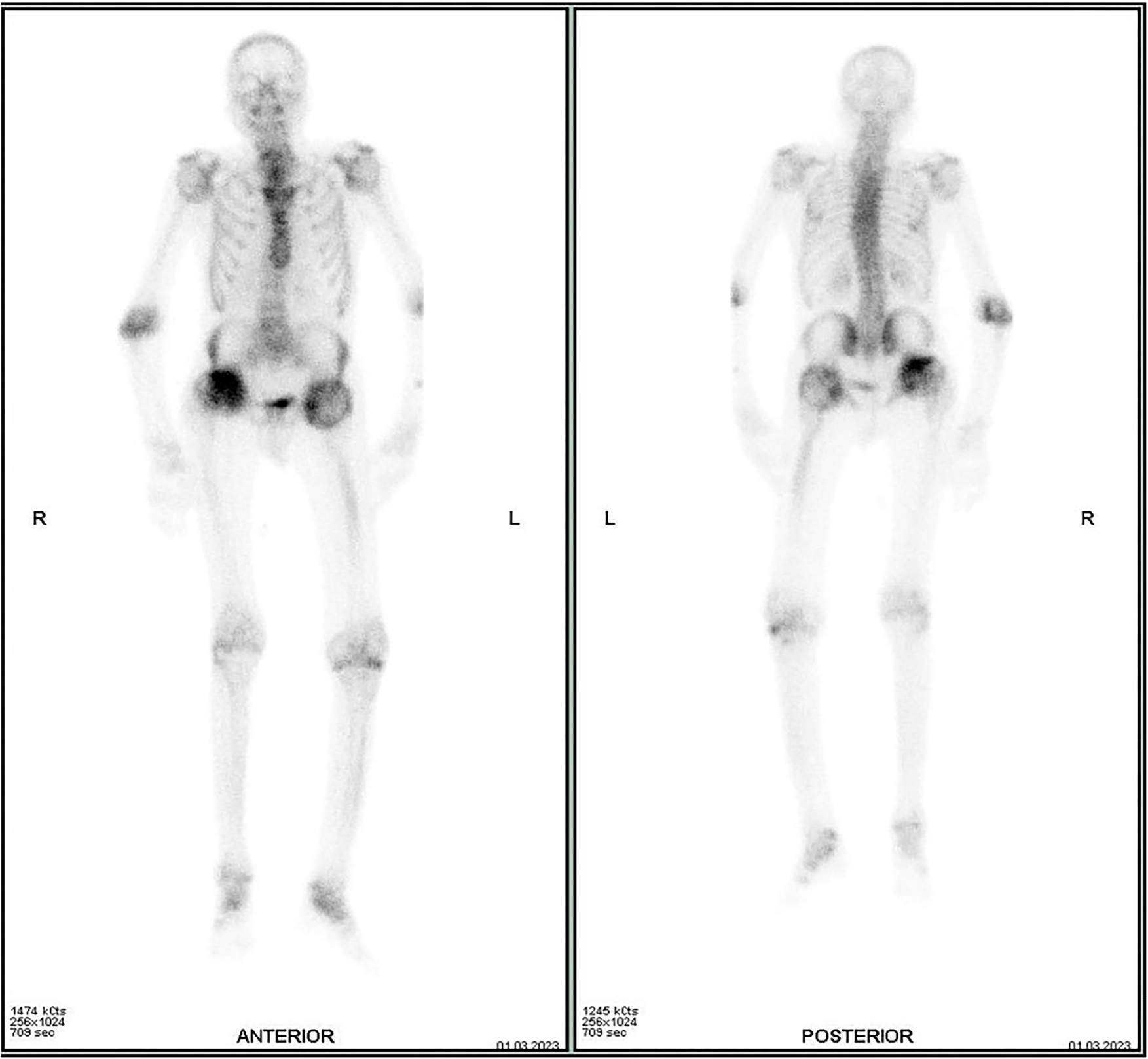
A bone scan with 99mTc-methylene diphosphonate 99mTc-(MDP), performed 3 hours after the intravenous injection of 666 MBq (18 mCi) of 99mTc-MDP, revealed heterogeneous and abnormally increased activity with deformities of the femoral heads, elbows, knees, and ankles, a short spine, and marked thoracolumbar scoliosis, consistent with polyostotic SEDT involvement. Focal and linear areas of increased uptake were seen in the knees and hips, corresponding to periarticular ossifications and bony bridges in the active phase (Figure 1).
SED is a subgroup of bone dysplasias that affects the spine and the epiphyses of long bones. It includes three major forms: congenital SED, late-onset (tarda) SED, and SED associated with progressive arthropathy (). More recently, at least four types of SEDT, some with autosomal recessive inheritance and others with autosomal dominant inheritance, have also been described (). The classical form of SEDT, as illustrated in this case, is a genetic disorder inherited in an X-linked recessive pattern, manifesting exclusively in males. It results from mutations in the SEDL gene located on Xp22.12-p22.31 and manifests around puberty with back pain, a short stature and a short trunk, while the extremities and face remain unaffected. Early onset degenerative joint disease, particularly affecting the spine and hips, usually develops in early adulthood. Radiologic abnormalities of the vertebral bodies include platyspondyly and a central hump (,). Bone scan is rarely indicated in SEDT; however, the skeletal involvement observed in this condition is unusual, requiring a thorough understanding of the distribution pattern of bone abnormalities for accurate interpretation and to avoid confusion with other entities, thereby ensuring appropriate management ().
Footnotes
Ethics
Informed Consent: Informed consent was obtained from the patient.
Authorship Contributions
Surgical and Medical Practices: I.Z., Concept: I.Z., S.O.N., M.A., A.D., Data Collection or Processing: I.Z., S.O.N., A.D., Analysis or Interpretation: I.Z., M.A., Literature Search: I.Z., S.O.N., M.A., Y.B., O.A.S., A.D., Writing: I.Z., S.O.N., A.D.
Conflict of Interest: No conflicts of interest were declared by the authors.
Financial Disclosure: The authors declare that this study has received no financial support.
References
Kocyigit H, Arkun R, Ozkinay F, Cogulu O, Hizli N, Memis A. Spondyloepiphyseal dysplasia tarda with progressive arthropathy. Clin Rheumatol. 2000;19:238-241.
Panda A, Gamanagatti S, Jana M, Gupta AK. Skeletal dysplasias: a radiographic approach and review of common non-lethal skeletal dysplasias. World J Radiol. 2014;6:808-825.
Gedeon AK, Tiller GE, Le Merrer M, Heuertz S, Tranebjaerg L, Chitayat D, Robertson S, Glass IA, Savarirayan R, Cole WG, Rimoin DL, Kousseff BG, Ohashi H, Zabel B, Munnich A, Gecz J, Mulley JC. The molecular basis of X-linked spondyloepiphyseal dysplasia tarda. Am J Hum Genet. 2001;68:1386-1397.
Yang F, Xu HQ, Li CL, Yang JG. Incidental finding of Tc-99m MDP bone scintigraphy in a case of X-linked spondyloepiphyseal dysplasia tarda. Clin Nucl Med. 2012;37:193-195.

