

Abstract
A 63-year-old woman presented to the emergency department with dyspnea, chest pain, and a palpable right breast mass. Non-contrast thoracic computed tomography (CT) scan revealed suspicious lesions in the right upper lung lobe and right breast, as well as right pleural effusion. For further evaluation, 18F-fluorodeoxyglucose (FDG) positron emission tomography (PET)/CT scan was performed. There was increased FDG uptake in the right breast, right lung, liver, bones, and lymph nodes. Histopathological assessment confirmed that the breast mass was a metastatic lesion originating from a primary small-cell lung carcinoma. The manifestation of primary lung carcinoma with a metastatic mass lesion in the breast region is a rare condition. The manifestation can lead to diagnostic challenges, particularly when distinguishing it from a primary breast tumor. 18F-FDG PET/CT may provide valuable information for staging and surveillance, especially in patients with atypical metastatic patterns. This case highlights that unexpected metastatic sites can significantly affect treatment strategies and are frequently associated with worse prognosis.
Keywords: Small cell lung carcinoma, breast mass, breast metastasis, 18F-FDG PET/CT
,
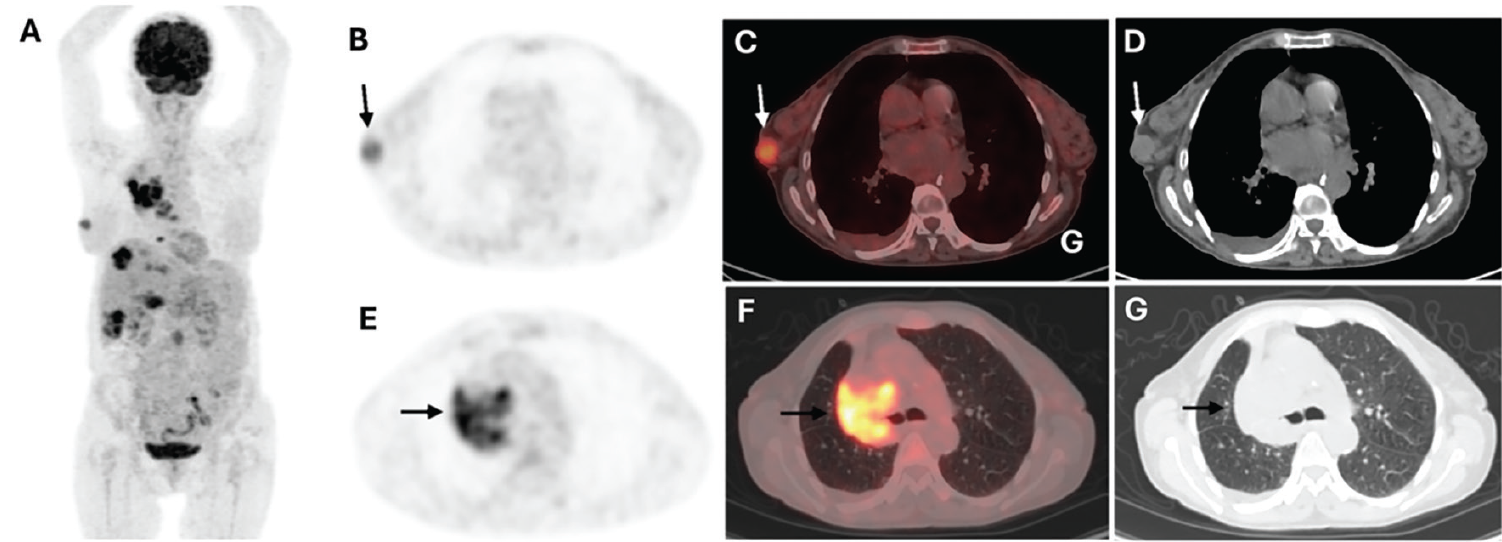
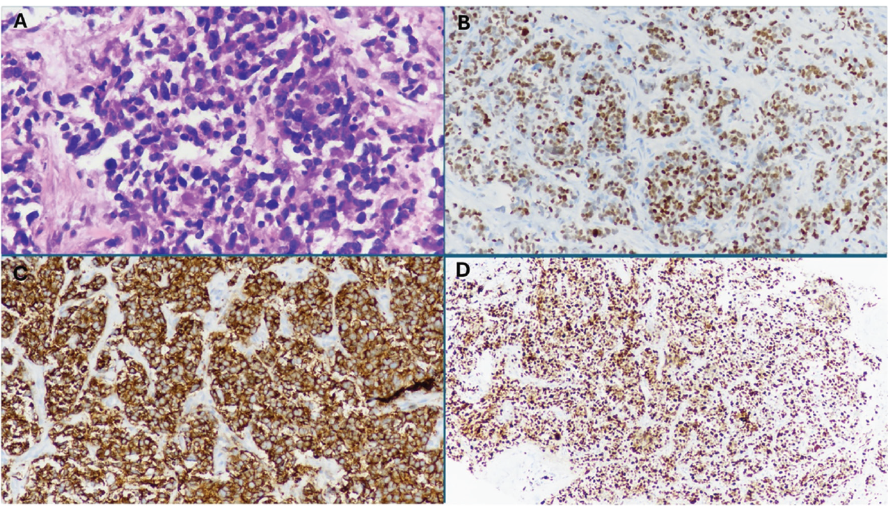
The extramammary metastases to the breast are an infrequent manifestation, and their prevalence has been reported to vary between 0.1% and 5.0% ().
The most common extramammary malignancies that metastasize to the breast are lymphoma, malignant melanoma, cancers of the lung, thyroid, gallbladder, cervix, and ovary (). Metastatic involvement of the breast from extramammary solid organ tumors represents a significant diagnostic challenge in routine clinical practice. Breast metastasis from lung cancer is an extremely rare clinical manifestation with an incidence of 0.2-1.3% (). It has been suggested that lung cancer cells can spread to the pleura, infiltrate the axillary lymph nodes, and then reach the ipsilateral breast via retrograde lymphatic pathways ().
Breast metastases from non-SCLC (NSCLC) have been reported more frequently than those from SCLC metastases (). However, SCLC metastases to the breasts have occurred synchronously. It may be that SCLC is a very aggressive entity and is characterized by rapid tumor growth. Most patients have widespread metastases known as extensive-stage SCLC at the time of initial diagnosis (). The most common metastatic sites of SCLC are mediastinal lymph nodes, liver, bone, and brain, respectively. It has been reported that the median overall survival for extensive-stage SCLC is approximately 12 months, while the 5-year survival rate for all SCLC patients remains only 6.8% (). Furthermore, the median survival time for advanced-stage breast cancer patients with lung metastases has been reported to be approximately 21 months (). An accurate diagnosis of breast mass is crucial in determining appropriate treatment strategies and improving patient outcomes. However, the clinical presentation of breast metastases concurrent with primary lung carcinoma is extremely rare and can complicate differential diagnosis. Consequently, in patients without a prior history of malignancy who present with a breast mass, both primary breast cancer and extramammary metastasis should be considered, as demonstrated in this case.
Footnotes
Ethics
Informed Consent: Informed consent was obtained from the patient.
Authorship Contributions
Surgical and Medical Practices: H.T.K., A.T.A., Concept: N.A.D., Ö.Ö., Design: N.A.D., D.Ç, Ö.Ö., Data Collection or Processing: D.Ç., H.T.K., A.T.K., Analysis or Interpretation: N.A.D, D.Ç., Literature Search: N.A.D., Ö.Ö., Writing: N.A.D D.Ç.
Conflict of Interest: No conflicts of interest were declared by the authors.
Financial Disclosure: The authors declare that this study has received no financial support.
References
Li J, Wahab R, Brown AL, Guarnieri B, Lewis K, Mahoney MC, Vijapura C. Extramammary metastases to the breast. Radiographics. 2023;43:e230036.
Zhou P, Chang N, Abraham SC, Albarracin CT, Huo L, Chen H, Ding Q, Resetkova E, Middleton LP, Sahin AA, Bu H, Wu Y. Metastatic nonhematopoietic neoplasms to the breast: a study of 238 cases. Hum Pathol. 2022;125:59-67.
Lee AH. The histological diagnosis of metastases to the breast from extramammary malignancies. J Clin Pathol. 2007;60:1333-1341.
Malek D, Buccheri S, Dey CB, Samli B, Plemmons J. Lung cancer metastasis to the breast mimicking inflammatory breast carcinoma on imaging. Radiol Case Rep. 2019;14:1500-1505.
Zhu Y, Liu WW, Wu Q, Yao JH, Zhou ZG, Yang Y. Clinical and molecular characteristics of secondary breast metastases from primary lung cancer: a study of 22 Chinese cases. Int J Clin Exp Pathol. 2020;13:1880-1885.
Ganti AKP, Loo BW, Bassetti M, Blakely C, Chiang A, D’Amico TA, D’Avella C, Dowlati A, Downey RJ, Edelman M, Florsheim C, Gold KA, Goldman JW, Grecula JC, Hann C, Iams W, Iyengar P, Kelly K, Khalil M, Koczywas M, Merritt RE, Mohindra N, Molina J, Moran C, Pokharel S, Puri S, Qin A, Rusthoven C, Sands J, Santana-Davila R, Shafique M, Waqar SN, Gregory KM, Hughes M. Small cell lung cancer, version 2.2022, NCCN Clinical Practice Guidelines in Oncology. J Natl Compr Canc Netw. 2021;19:1441-1464.
Cittolin-Santos GF, Knapp B, Ganesh B, Gao F, Waqar S, Stinchcombe TE, Govindan R, Morgensztern D. The changing landscape of small cell lung cancer. Cancer. 2024;130:2453-2461.
Xiao W, Zheng S, Liu P, Zou Y, Xie X, Yu P, Tang H, Xie X. Risk factors and survival outcomes in patients with breast cancer and lung metastasis: a population-based study. Cancer Med. 2018;7:922-930.

