

Abstract
The majority of metastatic pleural lesions are caused by malignancies such as bronchogenic carcinoma (40%), breast cancer (20%), lymphoma (10%), and ovarian or gastric carcinomas (5%). However, pleural metastases from thymoma are extremely rare. In this report, we present the 18F-fluorodeoxyglucose (18F-FDG) positron emission tomography/computed tomography (PET/CT) imaging findings of a patient with thymoma and extensive pleural metastases. Although biopsy remains the gold standard for diagnosis, it is important to consider high grade thymoma in the differential diagnosis, as extensive pleural involvement observed on 18F-FDG PET/CT imaging can mimic both primary and metastatic pleural malignancies. Recognizing this possibility can assist in more accurate interpretation of imaging findings.
Keywords: Thymoma, pleura, metastasis, 18FDG, PET/CT
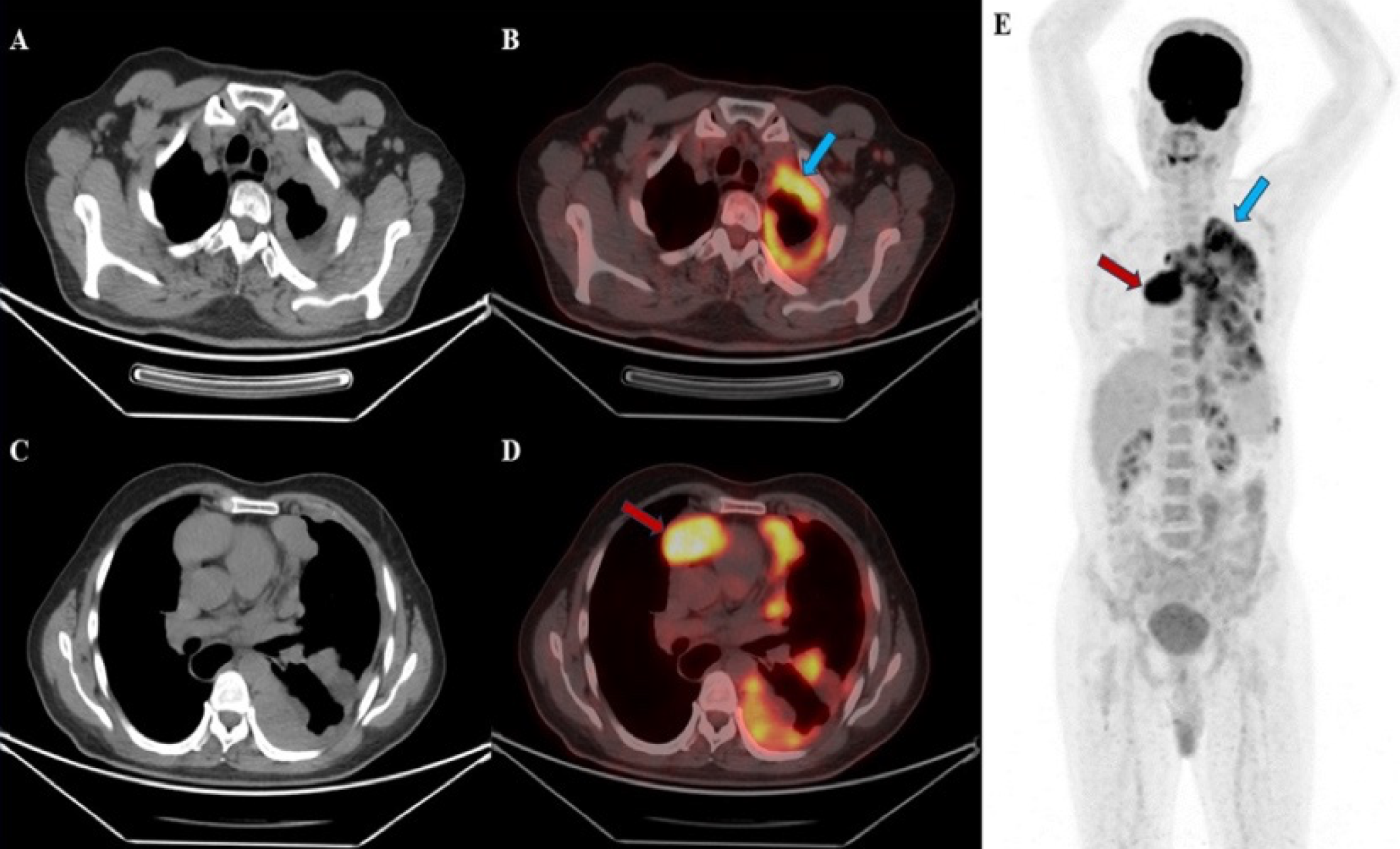
18F-fluorodeoxyglucose (18F-FDG) positron emission tomography (PET))/CT imaging provided further diagnostic confirmation, revealing increased 18F-FDG uptake in a soft tissue lesion located in the anterior mediastinum measuring 64 × 44 mm (Figures D, E, red arrow) with a maximum standardized uptake value (SUVmax) of 11.11. Additional elevated FDG uptake was observed in pleural masses in the left hemithorax (Figures B, D, blue arrow) with an SUVmax of 7.99. No pleural effusion was identified, and physiological FDG distribution was noted in other regions of the body (Figure E).
Thymomas are classified into five histological subtypes (A, AB, B1, B2, and B3) based on the WHO classification. Subtypes A, AB, and B1 are considered low risk for recurrence, whereas subtypes B2 and B3 are categorized as high-risk, with a greater propensity for metastasis and recurrence (). Thymomas are rare neoplasms that generally grow slowly and exhibit local spread. Common metastatic sites include the pleura, pericardium, and diaphragm, while extrathoracic metastases are uncommon ().
Although pleural metastases of high-risk thymoma are rare, they can mimic the imaging features of primary pleural malignancies or metastatic pleural disease (). Therefore, it is important to consider high-grade thymoma in the differential diagnosis when pleural involvement is observed. Careful evaluation of the anterior mediastinum in such cases may facilitate accurate diagnosis and significantly impact treatment planning. This case highlights the diagnostic value of 18F-FDG PET/CT and underscores its critical role in differentiating thymomas with pleural involvement (,).
Footnotes
Ethics
Informed Consent: The patient consent was obtained.
Authorship Contributions
Surgical and Medical Practices: M.O.K., B.O.T., Concept: M.O.K., B.O.T., Design: M.O.K., B.O.T., Data Collection or Processing: M.O.K., B.O.T., Analysis or Interpretation: M.O.K., B.O.T., Literature Search: M.O.K., B.O.T., Writing: M.O.K., B.O.T.
Conflict of Interest: No conflicts of interest were declared by the authors.
Financial Disclosure: The authors declare that this study has received no financial support.
References
Sandri A, Cusumano G, Lococo F, Alifano M, Granone P, Margaritora S, Cesario A, Oliaro A, Filosso P, Regnard JF, Ruffini E. Long-term results after treatment for recurrent thymoma: a multicenter analysis. J Thorac Oncol. 2014;9:1796-1804.
Falkson CB, Bezjak A, Darling G, Gregg R, Malthaner R, Maziak DE, Yu E, Smith CA, McNair S, Ung YC, Evans WK; Lung Cancer Disease Site Group of Cancer Care Ontario’s Program in Evidence-Based Care. The management of thymoma: a systematic review and practice guideline. J Thorac Oncol. 2009;4:911-919.
Qureshi NR, Gleeson FV. Imaging of pleural disease. Clin Chest Med. 2006;27:193-213.
Tumapon D, Kim EE. Recurrent Thymoma with lung, pleural, and splenic metastases demonstrated on F-18 FDG PET/CT. OALib. 2023;10:1-6.
Gajendra S, Sahoo MK. (18)F FDG-PET/CT of malignant thymoma with pleural and diaphragmatic metastases. Lung India. 2016;33:116-177.

