

Abstract
A 68-year-old man with progressive exertional dyspnea after an upper respiratory tract infection underwent contrast-enhanced thorax computed tomography (CT) to exclude pulmonary thromboembolism. The radiological findings suggested pneumonia or alveolar edema. 18F-Fluorodeoxyglucose positron emission tomography/CT (18F-FDG PET) scan was conducted due to progressive radiological findings. It revealed increased FDG uptake in the prostate gland, mediastinal, abdominopelvic multiple lymph nodes and bilateral lung lesions. 68Ga- prostate-specific membrane antigen-11 (PSMA) PET/CT scan was performed due to low FDG uptake in the lymph nodes and elevated plasma total prostate-specific antigen values. Moderate to high PSMA uptake corresponded to the localizations of FDG uptake on PET/CT. Furthermore, histopathological and immunohistochemical examinations demonstrated that the bilateral lung lesions and bilateral pleural effusion were compatible with metastases from prostate adenocarcinoma.
Keywords: Prostate cancer, 68Ga-PSMA PET/CT, pulmonary lymphangitic spread, pleural metastasis, pneumonia, 18F-FDG PET/CT
,
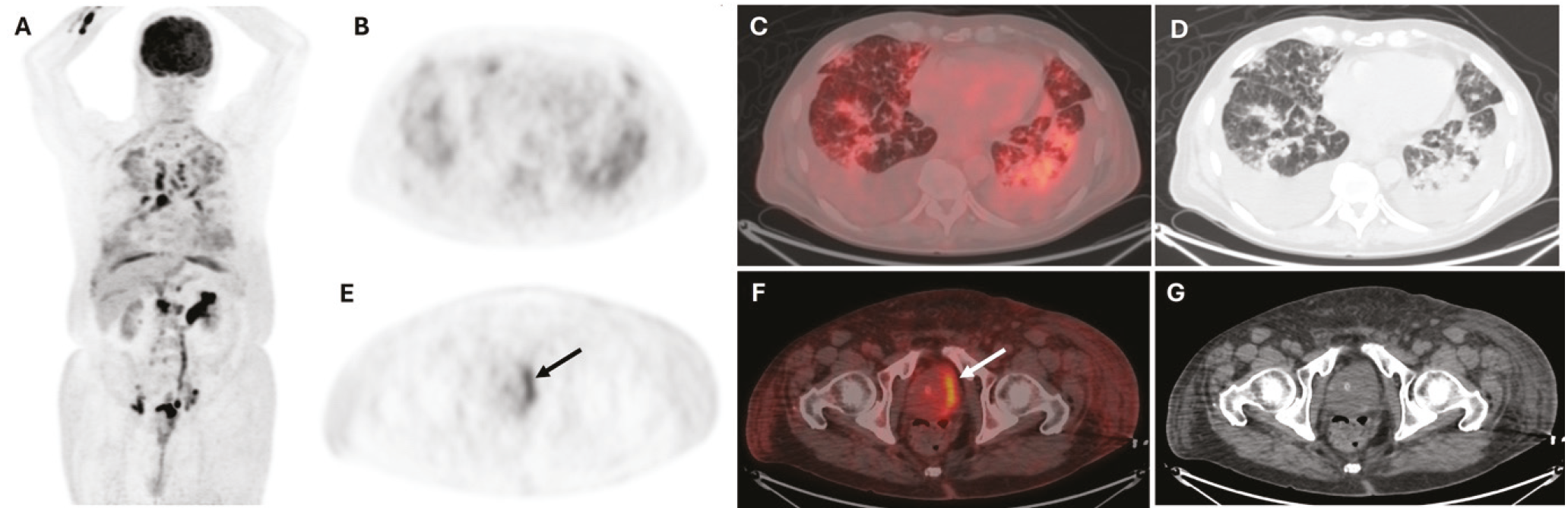
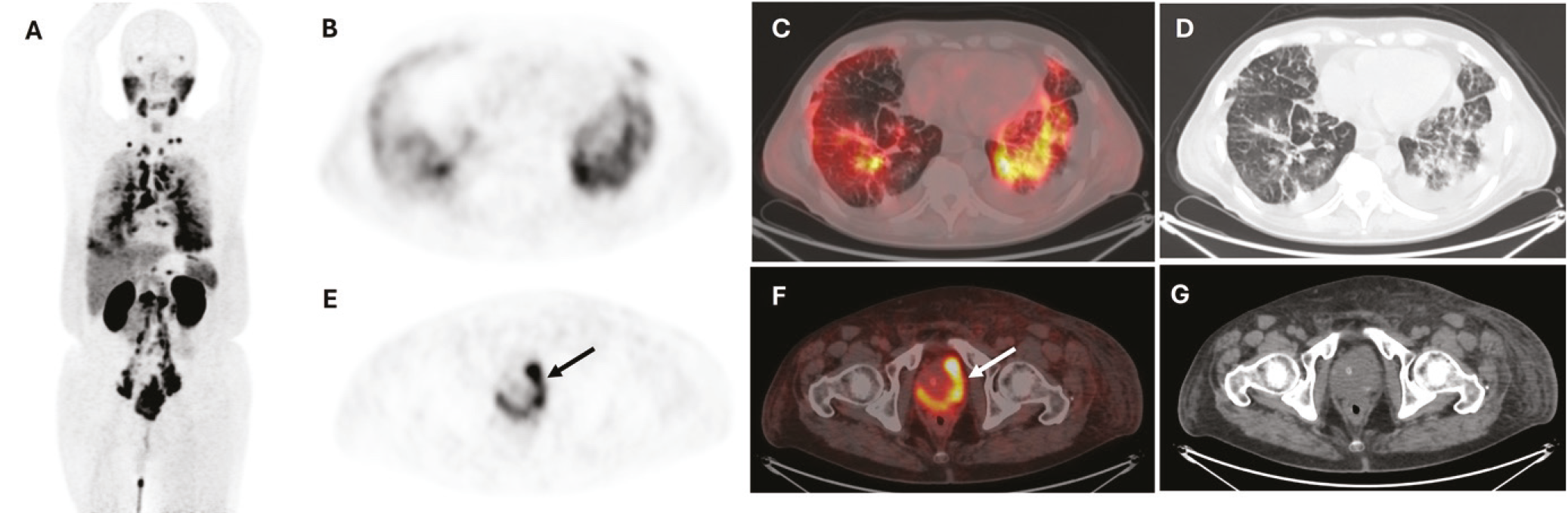
PSMA-ligand PET/CT plays a crucial role in detecting biochemical recurrence and staging intermediate- to high-risk PCa (). In the diagnosis and staging of primary PCa, the role of 18F-FDG PET/CT is considerably limited (,). Therefore, integrating PSMA-ligand PET/CT and 18F-FDG PET/CT can provide a more comprehensive assessment of metastatic disease, particularly in atypical presentations.
PCa may metastasize the lungs, most commonly presenting with nodular involvement. Pulmonary lymphangitic spread (PLC) with ground-glass opacities, bilateral malignant pleural effusions, and thickenings in PCa are exceedingly rare (,,). Pulmonary metastases from PCa may be overlooked on conventional imaging, particularly in patients with non-specific respiratory symptoms. Furthermore, both pulmonary metastases from PCa and certain benign or malignant lung diseases may demonstrate increased uptake of FDG and PSMA radiotracers ().
In this case, 18F-FDG PET/CT revealed pathological radiotracer uptake in the prostate gland preceding both symptomatic presentation and laboratory findings. However, 68Ga-PSMA PET/CT provided superior accuracy in determining the extent of disease spread, thereby enabling informed treatment planning. Therefore, it should always be considered that bilateral pleural effusion and PLC with ground-grass opacity may represent metastatic spread from PCa, even in patients without a prior history of malignancy.
Footnotes
Ethics
Informed Consent: Written informed consent was obtained from the patient.
Authorship Contributions
Surgical and Medical Practices: N.A.D., Concept: E.T., A.N.Ç., Ö.Ö., Data Collection or Processing: E.T., A.N.Ç., Analysis or Interpretation: N.A.D., Ö.Ö., Literature Search: A.N.Ç., B.B., Writing: N.A.D.
Conflict of Interest: No conflicts of interest were declared by the authors.
Financial Disclosure: The authors declare that this study has received no financial support.
References
Sachpekidis C, Alberts I, Rominger A, Afshar-Oromieh A. 68Ga-prostate-specific membrane antigen uptake in a malignant pleural effusion from metastatic prostate cancer after pleurodesis. Clin Nucl Med. 2019;44:838-839.
Jadvar H. Imaging evaluation of prostate cancer with 18F-fluorodeoxyglucose PET/CT: utility and limitations. Eur J Nucl Med Mol Imaging. 2013;40 (Suppl 1):S5-S10.
Makis W, Ciarallo A. Clinical significance of 18 F-fluorodeoxyglucose avid prostate gland incidentalomas on positron emission tomography/computed tomography. Mol Imaging Radionucl Ther. 2017;26:76-82.
Skrobisz K, Miszewski K, Miszewska L, Bieńkowski M, Matuszewski M, Studniarek M. Pleural metastasis as an initial presentation of prostate cancer: case report and literature review. Diagnostics. 2025;15:666.
Hibino M, Maeda K, Horiuchi S, Fukuda M, Kondo T. Pulmonary lymphangitic carcinomatosis with ground-glass opacities as presentation of prostate cancer. Respirol Case Rep. 2018;6:e00347.
Knight JC, Ray MA, Benzaquen S. Malignant pleural effusion from prostate adenocarcinoma. Respir Med Case Rep. 2014;13:24-25.
Srinivasan R, Cook GJR, Patel N, Subesinghe M. Prostate specific membrane antigen (PSMA) avid nonprostatic benign and malignant disease: a pictorial review. Clin Radiol. 2024;79:639-656.

