

Abstract
An 82-year-old patient with high-risk prostate adenocarcinoma, previously treated with radiotherapy and hormone therapy, presented with biochemical recurrence, as evidenced by a prostate-specific antigen level of 10 ng/mL. A positron emission tomography/computed tomography (PET/CT) scan using 18F-prostate-specific membrane antigen (PSMA ) 1007 revealed significant uptake at the prostate apex and multiple hypermetabolic osseous lesions in the sternum and L3, suggestive of local recurrence and metastasis. However, several hypermetabolic foci in the abdominopelvic region raised suspicion for potential involvement of the lumbar-aortic lymph nodes due to their moderate to intense uptake. After further investigation, these findings were attributed to active aortic calcifications. This case highlights a rare cause of false-positive results in 18F-PSMA 1007 PET/CT imaging and underscores the need for additional evaluations, such as abdominopelvic magnetic resonance imaging and renal and phosphocalcic assessments, when such results are suspected.
Keywords: Aortic calcifications, lymph nodes, 18F-PSMA1007
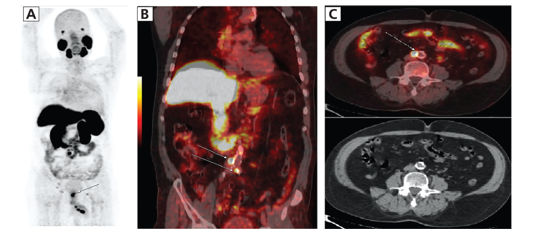
An 82-year-old man was diagnosed with high-risk prostate cancer (PCa) in 2020 and treated with radiotherapy and hormone therapy. In 2024 the patient had a biochemical recurrence with a prostate-specific antigen (PSA) measured at 10 ng/mL. A PET/CT scan with 18F-PSMA 1007 showed significant uptake at the prostate apex (SUVmax=12.9), indicating local recurrence (Figure A arrow). In addition, multiple hypermetabolic osseous lesions were identified in the sternum (SUVmax=4.2) and L3 (SUVmax=4.7), suggesting osseous metastasis. However, several hypermetabolic foci were also noted on the MIP in the abdominopelvic region (Figure A) raising initial concerns about potential involvement of lumbar-aortic lymph nodes due to the moderate to intensive uptake. These findings were attributed to active aortic calcifications (SUVmax=3.3-19.5) (Figures B and C arrows). This aspect was assessed by an abdominopelvic magnetic resonance imaging (MRI), which did not reveal any suspicious lymphadenopathy in the lumbo-aortic region. Additionally, an assessment of calcium-phosphorus balance showed a slightly elevated calcium at 110 mg/dL (normal value: 90-105 mg/dL), while renal function remained normal.
The application of 18F-PSMA-1007 PET/CT in imaging of PCa has significantly improved the detection of PSA relapse and metastasis, especially in patients with high-risk disease (,). However, false-positive findings may occur in several cases. Indeed, PSMA is highly expressed in the neovasculature of tumors, both benign and malignant. It plays a regulatory role in both angiogenesis and the expression of vascular growth factors, which are crucial for tumor growth and metastasis (). This expression is absent in endothelial cells of normal tissues but present in endothelial cells of tumors, leading to PSMA ligand uptake. This allows for the detection of tumor neovasculature in lesions, which can be problematic in non-prostatic tumors, where false-positive findings may occur due to enhanced blood flow and angiogenesis associated with tumor growth. Additionally, inflammation and infection can lead to PSMA expression in immune cells, although the precise mechanisms are not fully understood. Inflammatory conditions, particularly those associated with tissue remodeling and neovascularization, can increase the availability of PSMA ligands at the inflamed site (). Increased vascular permeability and regional blood flow at sites of infection or inflammation can facilitate PSMA ligand uptake, resulting in false-positive findings on PET scans (). Galiza Barbosa () demonstrated that increased blood flow in inflammatory tissues can lead to misinterpretation of PSMA-PET scans. The uptake of PSMA ligands by calcified vascular structures, such as those in the aorta, can mimic the appearance of malignant lesions on PET scans. Active aortic calcifications can result in elevated SUVmax values, as seen in our patient, leading to a mistaken impression of metastatic disease (). Such findings can be particularly misleading in elderly patients, who are more likely to have atherosclerotic changes and vascular calcifications. Islam et al. () further reinforce the importance of recognizing this limitation in PSMA PET/CT imaging. They recommend integrating other imaging modalities, such as CT or MRI, to better characterize ambiguous findings on PET/CT scans. By combining these modalities, clinicians can achieve a more accurate diagnosis, minimizing the risk of false-positive interpretations that could lead to unnecessary biopsies or other invasive procedures ().
Footnotes
Ethics
Informed Consent: The institutional review board of our institute “Med V Military Teaching Hospital”, approved this publication, and the requirement to obtain informed consent was waived.
Authorship Contributions
Surgical and Medical Practices: S.N.O., Concept: S.N.O., O.A.S., A.D., Design: S.N.O., O.A.S., I.Z., Data Collection or Processing: S.N.O., O.A.S., I.Z., M.A., Analysis or Interpretation: S.N.O., I.Z., M.A., Y.B., A.D., Literature Search: S.N.O., M.A., Writing: S.N.O.
Conflict of Interest: No conflicts of interest were declared by the authors.
Financial Disclosure: The authors declare that this study has received no financial support.
References
Afshar-Oromieh A, Babich JW, Kratochwil C, Giesel FL, Eisenhut M, Kopka K, Haberkorn U. The rise of PSMA ligands for diagnosis and therapy of prostate cancer. J Nucl Med. 2016;57:79S-89S.
Yordanova A. The role of PSMA PET/CT in prostate cancer: imaging and beyond. Cancer Letters. 2016;382:205-213.
Orevi M, Ben-Haim S, Abourbeh G, Chicheportiche A, Mishani E, Yutkin V, Gofrit ON. False positive findings of [18F]PSMA-1007 PET/CT in patients after radical prostatectomy with undetectable serum PSA Levels. Front Surg. 2022;9:943760.
Moukaddam H, El Saheb G, Omran N, El Ghawi N, Abi Ghanem A, Haidar M. Case report: positive pitfalls of PSMA PET/CT: diagnostic challenges in degenerative bone lesions including MODIC type 1. Front Nucl Med. 2024;4:1451848.
Galiza Barbosa F, Queiroz MA, Nunes RF, Bezerra ROF, Dalaqua M, Gustavo Rocha Resende R, Danil Faria Barbosa Lima E, Kobayashi M, Marins T, Santucci DF, Baroni RH. Nonprostatic diseases on PSMA PET imaging: a spectrum of benign and malignant findings. Cancer Imaging. 2020;20:23.
Islam R, Desai S, Moran M, Golombos DM. The role of PSMA PET imaging in prostate cancer: current applications and future directions. Curr Urol Rep. 2025;26:46.
Zaman MU, Fatima N, Zaman A, Sajid M, Zaman U, Zaman S. Diagnostic challenges in prostate cancer and 68Ga-PSMA PET imaging: a game changer? Asian Pac J Cancer Prev. 2017;18:2625-2628.

