

Abstract
Distant metastases from well-differentiated thyroid carcinoma typically involve the lungs and bones. We report a rare case of metastatic papillary thyroid carcinoma presenting as soft tissue lesion, detected by Iodine-131 whole-body scintigraphy in a 44-year-old asymptomatic male. The patient had previously undergone a total thyroidectomy and bilateral modified radical neck dissection, receiving radioiodine for pT3bN1bMx multifocal classical papillary thyroid carcinoma. Further evaluation with 18F-fluorodeoxyglucose positron emission tomography/computed tomography revealed additional sites of soft tissue and muscle metastasis, along with extensive nodal, lung, and skeletal involvement. These findings indicated advanced disease with a poor prognosis, necessitating a change in the treatment plan.
Keywords: Iodine-131, thyroid cancer, 18F-fluorodeoxyglucose positron emission tomography/computed tomography, soft tissue metastasis, muscle metastasis
,
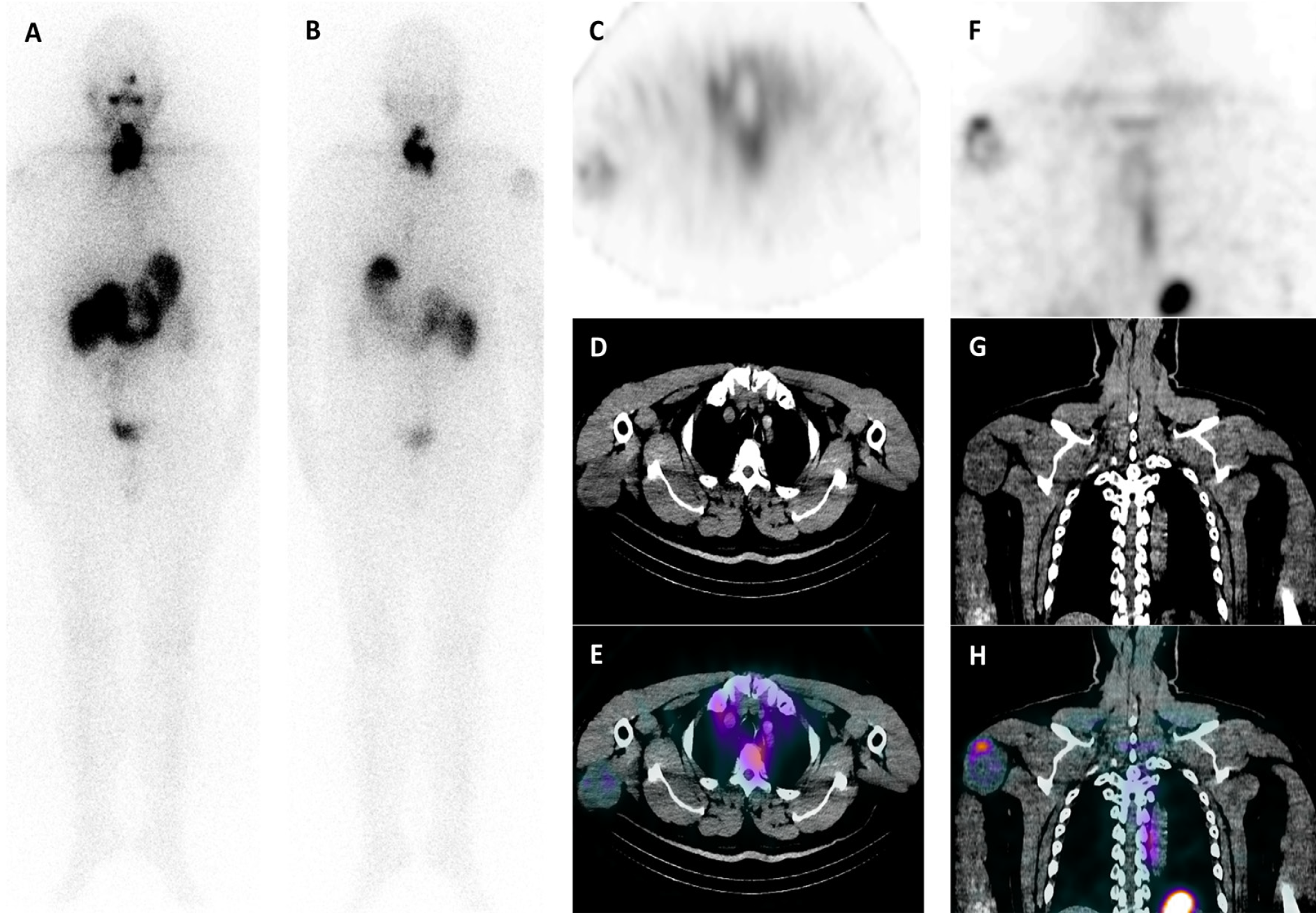
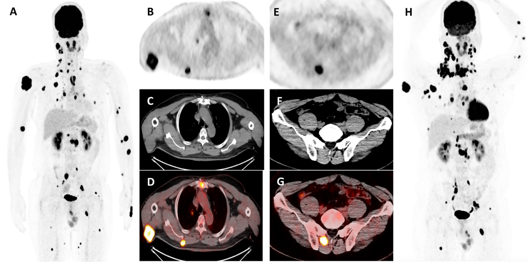
Distant metastases in differentiated thyroid carcinoma occur in approximately 5% to 10% of cases, typically affecting the lungs and bones (). Less common sites of dissemination, such as the liver, brain, soft tissue, and muscle, often indicate dedifferentiation of the disease and may arise years after the initial diagnosis (,). Soft tissue and muscle metastases from papillary thyroid carcinoma are rare and frequently identified incidentally on imaging due to the asymptomatic nature of most cases (,). These metastases generally signify advanced and widely disseminated disease, as in this case (,). However, there are reports of solitary soft tissue and muscle metastases in some patients (,,). Soft tissue and skeletal muscle are considered unfavorable environments for tumor proliferation due to factors such as anti-tumor cytokines (e.g., interleukin adenosine and leukemia inhibitory factor); the removal of lactic acid; mechanical disruption of tumor cells; inappropriate pH levels; variable blood flow; and continuous muscle motion (,,,,). Nevertheless, soft tissue and muscle metastasis can occur via multiple mechanisms, including intramuscular lymphatic spread, hematogenous dissemination, and perineural transmission (). In this context, 18F-FDG PET/CT is crucial when serum Tg levels are disproportionately high and do not correlate with Iodine-131 (131I) whole-body scan findings (). It helps identify iodine-refractory or dedifferentiated lesions, revealing occult metastases not seen on 131I imaging (,). In this case, the detection of soft tissue metastasis on 131I whole-body scintigraphy, along with extensive hypermetabolic 18F-FDG-avid lesions in unexpected sites, indicated a poor prognosis and shorter survival (,,,,), necessitating a change in treatment strategy.
Footnotes
Ethics
Informed Consent: The patient provided written informed consent.
Authorship Contributions
Surgical and Medical Practices: M.F.M.R., S.Z.A.H., Concept: M.F.M.R., S.Z.A.H., Design: M.F.M.R., S.Z.A.H., Data Collection or Processing: M.F.M.R., S.Z.A.H., Analysis or Interpretation: M.F.M.R., S.Z.A.H., Literature Search: M.F.M.R., S.Z.A.H., Writing: M.F.M.R., S.Z.A.H.
Conflict of Interest: No conflicts of interest were declared by the authors.
Financial Disclosure: The authors declare that this study has received no financial support.
References
Hitu L, Cainap C, Apostu D, Gabora K, Bonci EA, Badan M, Mester A, Piciu A. Skeletal muscle metastasis in papillary thyroid microcarcinoma evaluated by F18-FDG PET/CT. Diagnostics (Basel). 2020;10:100.
Califano I, Quildrian S, Coduti M, Rojas Bilbao E, Otero J, Califano L. Soft tissue metastases from differentiated thyroid cancer diagnosed by 18 F FDG PET-CT. Arq Bras Endocrinol Metabol. 2013;57:317-321.
Stephen J, Thomas MB, Thomas MB. A case of papillary thyroid cancer with soft tissue metastasis. Int Surg J. 2018;5:3425-3429.
Qiu ZL, Luo QY. Erector spinae metastases from differentiated thyroid cancer identified by I-131 SPECT/CT. Clin Nucl Med. 2009;34:137-140.
Ceriani L, Treglia G, Paone G, Bongiovanni M, Franscella S, Giovanella L. Unusual muscular metastases from papillary thyroid carcinoma detected by fluorine-18-fluorodeoxyglucose PET/MRI. J Clin Endocrinol Metab. 2013;98:2208-2209.
Tunio MA, Alasiri M, Riaz K, Alshakwer W, Alarifi M. Skeletal muscle metastasis as an initial presentation of follicular thyroid carcinoma: a case report and a review of the literature. Case Rep Endocrinol. 2013;2013:192573. Epub 2013 Mar 28.
Shin T, Hoang TD, Shane SP, Mai VQ, Shakir MKM. Multi‑focal metastatic papillary thyroid cancer to skeletal muscle with BRAFV600E mutation. Trends Med. 2020;20:2‑3.
Sarma M, Sonik B, Subramanyam P, Sundaram PS. Isolated skeletal muscle metastatic deposit in a patient with micropapillary carcinoma thyroid identified by 18F FDG PET CT. J Egypt Natl Canc Inst. 2015;27:47-50.
Herbowski L. Skeletal muscle metastases from papillary and follicular thyroid carcinomas: an extensive review of the literature. Oncol Lett. 2018;15:7083-7089.
Stergioula A, Pantelis E, Kormas T, Agrogiannis G. Case report: skeletal muscle metastasis from follicular thyroid carcinoma presenting as synovial sarcoma. Front Oncol. 2023;13:994729.

