

Abstract
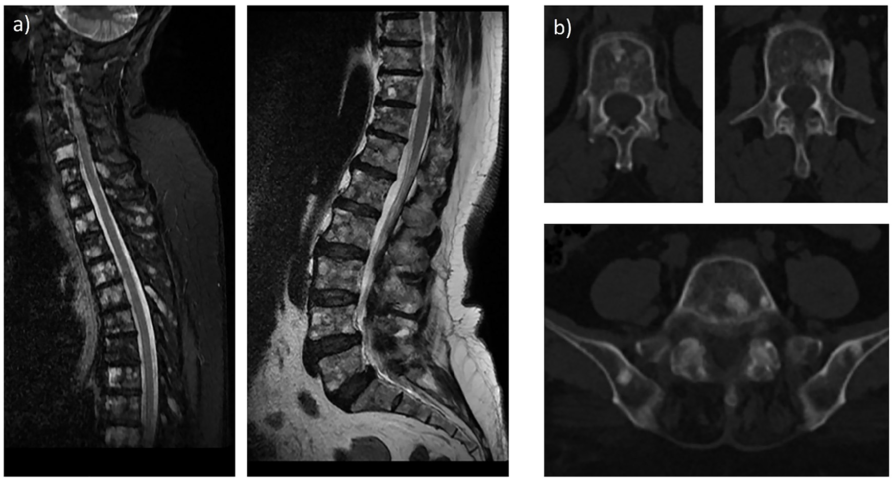
Identifying osseous metastases by imaging is essential and may be challenging in patients with lobular breast cancer. We present a case of a 66-year-old woman with lobular breast cancer who underwent 18F- fluorodeoxyglucose positron emission tomography/computed tomography (FDG PET/CT) for staging purposes. 18F-FDG PET/CT reveals minimal FDG uptake in the primary tumor cells. Prominent sclerotic lesions with low FDG avidity are seen in the spinal and pelvic bones. The subsequent Tc-99m methylene diphosphonate bone scan is unremarkable. The magnetic resonance imaging (MRI) reveals bone metastases. MRI may be beneficial in invasive lobular carcinoma. MRI facilitates improved metastatic evaluation, especially in bone-only and bone-predominant metastatic malignancies, when assessment with 18F-FDG PET/CT may be difficult and constrained.
Keywords: Lobular breast cancer, 18F-FDG PET/CT, osseous metastases, MRI
,
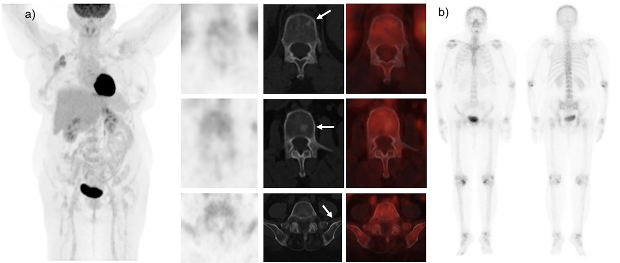
18F-fluorodeoxyglucose positron emission tomography/CT(FDG PET) plays a meaningful role in the staging and restaging of breast cancer. However, its sensitivity and detection rates are lower in lobular carcinoma, attributable to the lessened FDG-avidity of malignant cells (). ILC demonstrates lower maximum standardized uptake values standardized uptake value than invasive ductal carcinoma (IDC) on account of multiple mechanisms, such as lower tumor cell density, reduced proliferation rates, diffuse infiltration into encompassing tissues, and diminished GLUT-1 expression ().
In women with breast cancer, bone is the principal site of distant metastasis. It is also the primary site of metastasis in 50% of patients (). Individuals diagnosed with lobular breast cancer display a greater tendency for bone metastases compared to those with ductal malignancies and more often appear sclerotic on CT scans than those with IDC. 18F-FDG PET/CT is more sensitive in detecting lytic bone metastases in breast cancer patients, while Tc-99m methylene diphosphonate bone scintigraphy demonstrates greater sensitivity for detecting sclerotic bone metastases. Recently, a study by Usmani et al. () showed low sensitivity and precision of 18F-FDG PET/CT in identifying bone metastases in patients with lobular breast cancer. The absence of uptake on whole body bone scan despite the presence of sclerotic lesions presents an intriguing diagnostic phenomenon. This discordance may be attributed to several factors, including the subtle nature and small size of the sclerotic lesions, which could fall below the resolution threshold of bone scintigraphy. Additionally, these findings might represent very early metastatic disease that is currently undetectable. Another consideration is the possibility of low osteoblastic activity despite the sclerotic appearance on CT, suggesting a unique biological behavior of these metastatic lesions.
MRI demonstrates superior accuracy in the diagnosis of bone metastasis associated with lobular breast cancer. It detects bone metastases at an earlier stage than other imaging techniques. It can identify subtle changes in bone marrow that may not be visible on bone scan, 18F-FDG PET or CT scans. The rate of misdiagnosis associated with MRI in identifying bone metastasis in lobular breast cancer is comparatively low (). In untreated patients with ILC, non-FDG-avid sclerotic osseous metastases were found to be significantly more prevalent than in those with IDC. The diagnostic confidence is lower in 18F-FDG PET/CT in ILC when evaluating for osseous metastases and consideration given to performing a biopsy/MRI or obtaining follow-up imaging before excluding metastatic disease. Our case highlights the need for careful evaluation of any sclerotic lesion in lobular breast cancer patients, even if negative on 18F-FDG PET/CT, and to consider alternative imaging modalities or more specific PET tracers such as 18F-fluoroestradiol and 68Ga-fibroblast activation protein inhibitor-04 as alternative options for comprehensive evaluation of lobular breast cancer metastases (,). Furthermore, PET/MRI emerges as a promising hybrid modality that could enhance diagnostic accuracy in such cases. This hybrid technology demonstrates higher sensitivity for detecting bone metastases compared to PET/CT, making it particularly valuable in cases of ILC where metabolic activity is characteristically low. The integration of PET/MRI into the diagnostic algorithm for ILC could potentially bridge the current imaging gaps, though availability and cost considerations may influence its implementation in clinical practice.
Footnotes
Ethics
Informed Consent: The consent was obtained for image use.
Authorship Contributions
Surgical and Medical Practices: S.U., K.A.R., A.J., A.A.B., N.A., Concept: S.U., K.A.R., A.J., Design: S.U., V.V.J., S.K., Data Collection or Processing: S.U., K.A.R., A.J., A.A.B., P.D., V.V.J., S.K., N.A., Analysis or Interpretation: S.U., A.J., A.A.B., Literature Search: S.U., N.A., Writing: S.U., K.A.R., N.A.
Conflict of Interest: No conflicts of interest were declared by the authors.
Financial Disclosure: The authors declare that this study has received no financial support.
References
Hogan MP, Goldman DA, Dashevsky B, Riedl CC, Gönen M, Osborne JR, Jochelson M, Hudis C, Morrow M, Ulaner GA. Comparison of 18F-FDG PET/CT for systemic staging of newly diagnosed invasive lobular carcinoma versus invasive ductal carcinoma. J Nucl Med. 2015;56:1674-1680.
Bos R, van Der Hoeven JJ, van Der Wall E, van Der Groep P, van Diest PJ, Comans EF, Joshi U, Semenza GL, Hoekstra OS, Lammertsma AA, Molthoff CF. Biologic correlates of (18)fluorodeoxyglucose uptake in human breast cancer measured by positron emission tomography. J Clin Oncol. 2002;20:379-387.
Hamaoka T, Madewell JE, Podoloff DA, Hortobagyi GN, Ueno NT. Bone imaging in metastatic breast cancer. J Clin Oncol. 2004;22:2942-2953.
Usmani S, Al Riyami K, Jain A, Alajmi AA, AlBaimani K, Dumasig P, Al Busaidi A, Al Sukati R. Enhancing precision in bone metastasis diagnosis for lobular breast cancer: reassessing the role of 18 F-FDG PET/CT. Nucl Med Commun. 2024;45:858-864.
Rong Y, Ren H, Ding X. MRI and bone scintigraphy for breast cancer bone metastase: a meta-analysis. Open Med (Wars). 2019;14:317-323.
Sahin E, Kus T, Aytekin A, Uzun E, Elboga U, Yilmaz L, Cayirli YB, Okuyan M, Cimen V, Cimen U. 68 Ga-FAPI PET/CT as an alternative to 18 F-FDG PET/CT in the imaging of invasive lobular breast carcinoma. J Nucl Med. 2024;65:512-519.
Ulaner GA, Jhaveri K, Chandarlapaty S, Hatzoglou V, Riedl CC, Lewis JS, Mauguen A. Head-to-head evaluation of 18 F-FES and 18 F-FDG PET/CT in metastatic invasive lobular breast cancer. J Nucl Med. 2021;62:326-331.

