Truncation Artifact Presenting as Cropped Projections and Wedge Defect in Sinogram During Single-Photon Emission Computed Tomography
PDF

Interesting Image

Truncation Artifact Presenting as Cropped Projections and Wedge Defect in Sinogram During Single-Photon Emission Computed Tomography

Mol Imaging Radionucl Ther 2026;35(1):51-54 DOI: 10.4274/mirt.galenos.2025.58265
, ,
PDF
Permissions

Abstract

Truncation artifact during single-photon emission computed tomography occurs when some part of body, such as left ventricle in myocardial perfusion imaging, lies outside the field-of-view during image acquisition. Improper adjustment of axis of rotation of detectors regarding the patient on the scanning table is the main cause. Large or slim patient body habitus is reported as source of this artifact. However, there may be other sources including that in our case, in which the artifact was present in prone position but not in supine imaging. The appearance was one-sided incremental cropping of projections of one of detectors and a wedge-shaped or triangular defect in the corresponding sinogram. This finding may suggest a possible mechanical instability of the gantry during motion over the patient’s left side in prone imaging.

Keywords: Truncation artifact, single-photon emission computed tomography, myocardial perfusion imaging

,
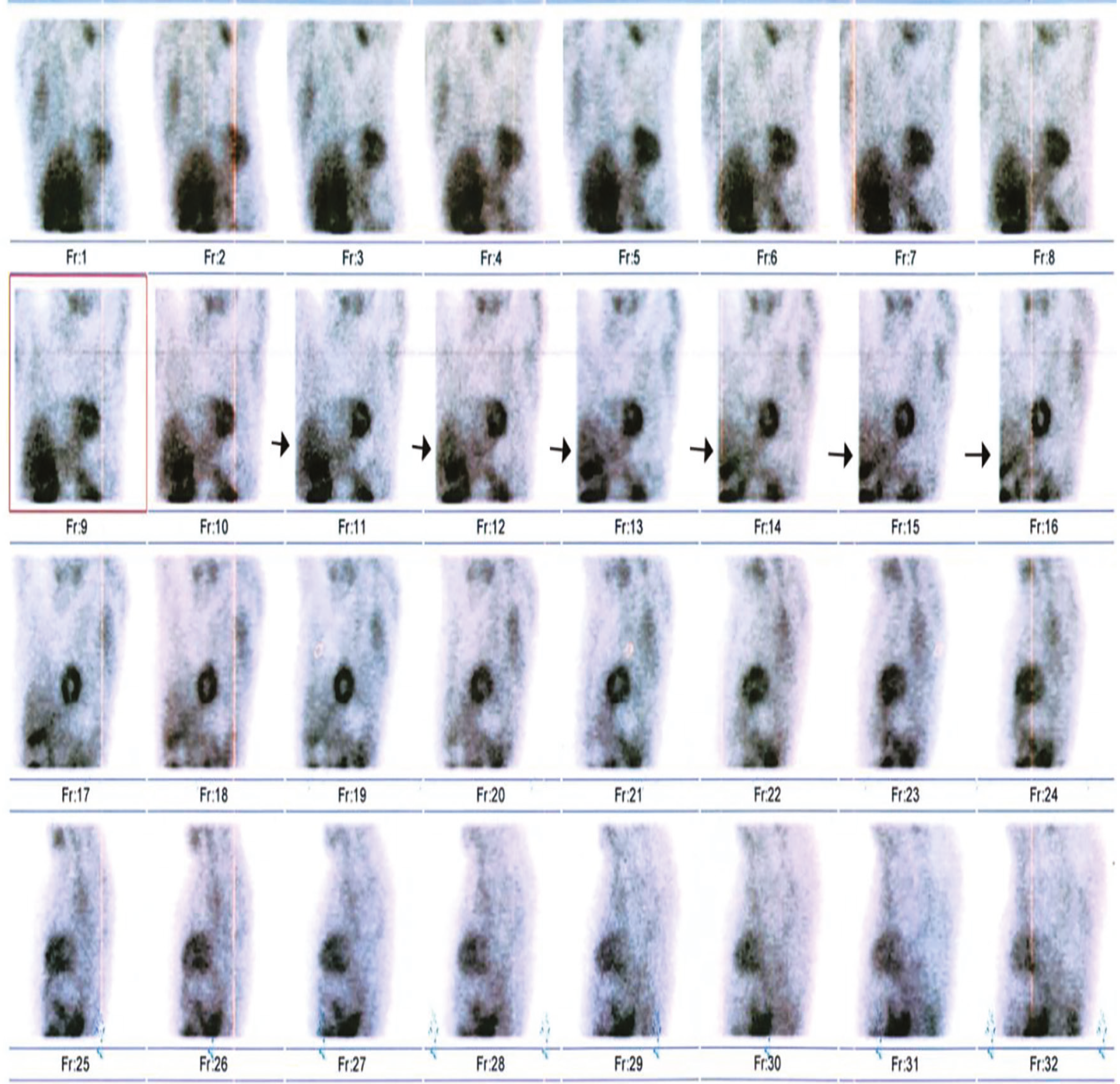
Figure 1
Figure 1. A 50-year-old man presented for myocardial perfusion single-photon emission computed tomography imaging. Scans are conducted during the stress and rest phases using a dual-headed gamma camera (Siemens Symbia T series) configured at a 90° angle. For resolving the diaphragmatic attenuation effect, prone imaging is performed. On prone imaging, left margin of projections 9 to 16 in the cinematic raw images, which include 32 projections in total and 16 projections for each detector, seems to be cropped incrementally in width.
View larger Download
Figure 2
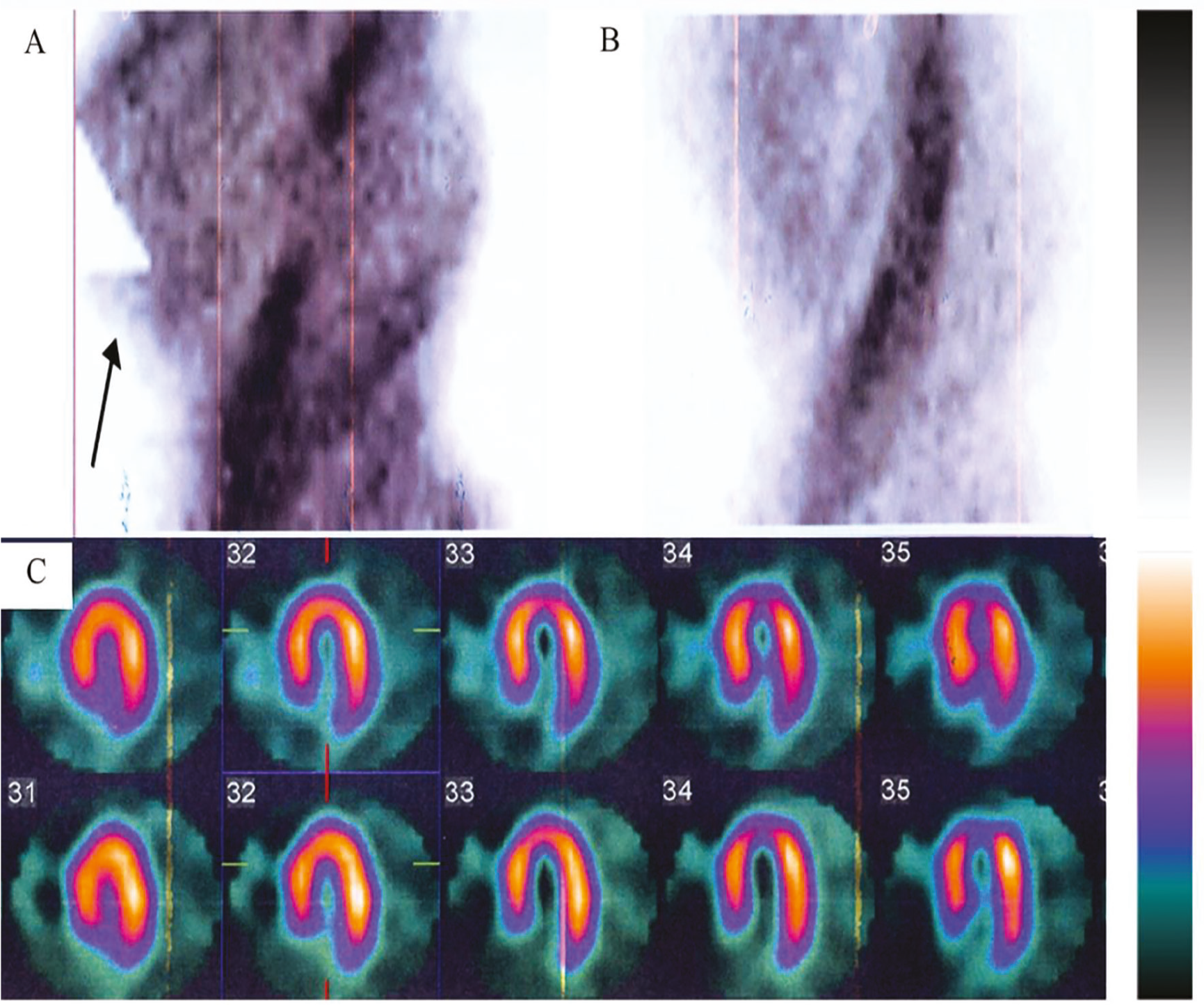
Figure 2. Inspecting the related sinograms, a wedge-shaped or triangular zone of absent activity was observed in the left margin of the sinogram of prone stress phase, (as shown by arrow in A) affecting several successive projections. The width of the defect is gradually increasing projection-by-projection until the end of the acquisition arc of detector 1, and, thereafter, abrupt resumption is seen at the beginning of the acquisition arc of detector 2. The tip of the triangular defect does not reach the sinusoidal band of left ventricle (LV) in the sinogram. Fortunately, the liver was resected, and LV remained unaffected. The sinogram of the rest phase is intact, (B). The tomographic slices (C) reconstructed from the horizontal long-axis of the prone and rest phases, using the iterative maximum likelihood expectation maximization method, from this projection dataset were free from this artifact. In rest and stress images performed in the supine position, no similar defect was observed in the sinogram and corresponding tomographic slices. The same finding was observed in other patients’ scans, mostly was acquired in prone position, and rarely in supine position (the latter in heavy patients). All routine quality control tests, including the center of rotation test, were acceptable. Truncation artifacts are one of several artifacts affecting single photon emission computed tomography imaging. When the LV is truncated, perfusion defects can occur. In rotating or cinematic images, this problem is easily recognizable, where in some successive projections, LV is abutting the border of projection or being cut. The solution to this problem is easy. When it is noticed, the operator should reposition the axis of rotation of the gantry (detector heads) to the longitudinal or z-axis of the patient. Another source may be encountered when imaging is performed using the option of auto-body contouring. In patients with small body size, some interference occurs between the two detectors because each detector tries to approach the patient. Using bags of saline or covering the patient with folded blankets may rectify this problem; otherwise, another orbit (circular or elliptical) should be applied (,,,,). In our case, none of the above sources was the culprit for this artifact. As mentioned, it arises mostly in prone imaging, where the detectors are positioned below the scanning table and disappears when imaging is done in the supine position. In the prone patient position, detectors are rotating around the patient while ascending above the scanning table. One plausible explanation is that because of mechanical instability, the lower positioned detector bears the weight of another detector and thus prevents proper positioning. This is responsible for the graded or incremental truncation of projections, creating a wedge-shaped appearance. The problem remained unresolved after repeated examination by the service engineers accredited by the vendor. This problem can be of interest to engineers and technicians. Fortunately, in this case, the heart was not affected, and the need for repeating or discarding the prone imaging was obviated.
View larger Download

Footnotes

Ethics

Informed Consent: An informed written consent was obtained from the patient both for performing the scanning and also for use and anonymous publishing of the images.

Authorship Contributions

Concept: M.Q., R.A., Design: M.Q., R.A., A.A., Data Collection or Processing: R.A., Analysis or Interpretation: M.Q., R.A., A.A., Literature Search: R.A., A.A., Writing: M.Q., R.A., A.A.

Conflict of Interest: No conflicts of interest were declared by the authors.

Financial Disclosure: The authors declare that this study has received no financial support.

References

1

Wosnitzer B, Gadiraju R, Depuey G. The truncation artifact. J Nucl Cardiol. 2011;18:187-191.

2

Matsumoto N, Suzuki Y, Yoda S, Hirayama A. The truncation artefact in patients with a high body mass index on myocardial perfusion SPECT. BMJ Case Rep. 2014;2014.

3

McGowan SE, Greaves CD, Evans S. An investigation into truncation artefacts experienced in cardiac imaging using a dedicated cardiac SPECT gamma camera with transmission attenuation correction. Nucl Med Commun. 2012;33:1287-1291.

4

Tsougos I, Alexiou S, Theodorou K, Valotassiou V, Georgoulias P. The prevalence of a false-positive myocardial perfusion stress SPET test in a skinny patient, induced by projection truncation. Hell J Nucl Med. 2015;18:79-80.

5

Yapici O, Baris S, Alic T, Basoglu T. Auto-contouring at 90 degrees dual head fitting angle: a potential cause of a myocardial perfusion SPET artifact in slim patients. Hell J Nucl Med. 2009;12:289-290.