

Abstract
A 49-year-old premenopausal woman with a history of left modified radical mastectomy and axillary lymph node dissection for invasive lobular carcinoma three years ago, underwent 18F-fluorodeoxyglucose positron emission tomography/computed tomography (18F-FDG PET/CT) due to gradually increasing CA15-9 and CEA levels during routine follow-up. Compared with previous 18F-FDG PET/CT images, the left ovary in the current images showed increased size and radiotracer uptake. Subsequently, total abdominal hysterectomy and bilateral salpingo-oophorectomy were performed, and histopathological evaluation confirmed unilateral metastasis in the left ovary due to primary breast cancer. Although rare, possible isolated ovarian metastasis should be considered in patients with breast cancer who have increased size and radiotracer uptake in ovarian tissue on follow-up 18F-FDG PET/CT scans.
Keywords: Breast carcinoma, 18F-FDG PET/CT, unilateral ovarian metastasis, isolated ovarian metastasis, ovarian uptake
,
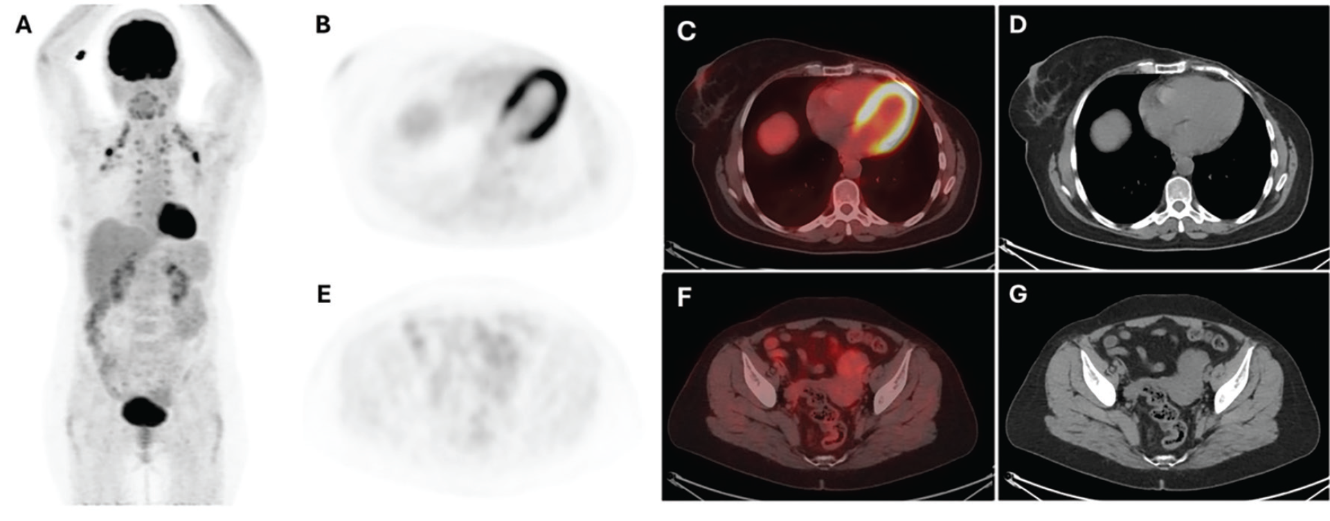
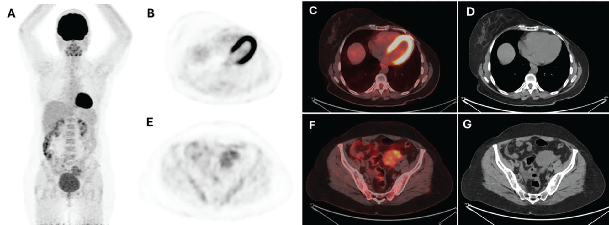
BC can frequently metastasize to bones, lungs, liver, and brain. However, invasive lobular carcinoma (ILC) has a greater tendency to metastasize to disseminated sites including the gastrointestinal (GI) tract, peritoneum, gynecological organs, skin, and adrenal glands (). Among gynecologic organs, ovaries are the most frequently site of metastasis of BC (). Tumors may metastasize to the ovaries through various routes, including direct, hematogenous, and lymphatic spread, as well as transcoelomic dissemination (). Several studies have shown that GI tract (gastric, colon and rectum) tumors are the most common primary neoplasm metastasizing to the ovary, followed by uterine, BC, pancreatic cancer, lymphoma, malignant melanoma and others (,).
Ovarian metastasis from BC is more frequently seen in younger and premenopausal women. They are asymptomatic until they reach a certain size, so they are frequently associated with other poor prognostic factors such as the presence of other metastatic sites, large primary breast lesion size, inflammatory BC, lymph node involvement, advanced stage (stage III-IV), and bilateralism (). Therefore, it is frequently associated with worse prognosis and outcomes, and a shorter 5-year survival rate (). Isolated unilateral ovarian metastasis from ILC is extremely rare. Furthermore, ILC shows low FDG uptake, so 18F-FDG PET/CT may have a lower impact on systemic staging for ILC than for invasive ductal carcinoma (). However, in this case, 18F-FDG PET/CT detected an isolated ovarian radiotracer uptake, raising suspicion of metastasis. This case report demonstrated that the presence of increased size and FDG uptake, merely in the isolated unilateral ovary, should be considered a potential indication of metastatic spread of BC.
Footnotes
Ethics
Informed Consent: The informed consent was obtained from the patient.
Authorship Contributions
Surgical and Medical Practices: N.A.D., Concept: E.T., Design: D.Ç., Data Collection or Processing: Ö.Ö., Analysis or Interpretation: D.Ç., Ö.Ö., Literature Search: E.T., Writing: N.A.D.
Conflict of Interest: No conflicts of interest were declared by the authors.
Financial Disclosure: The authors declare that this study has received no financial support.
References
DiPiro PJ, Tirumani SH, Cruz GP, Ramaiya NH, Lester SC, Shinagare AB. Lobular breast cancer: patterns of intraabdominal metastatic spread on imaging and prognostic significance. Abdom Radiol (NY). 2019;44:362-369.
Kutasovic JR, McCart Reed AE, Males R, Sim S, Saunus JM, Dalley A, McEvoy CR, Dedina L, Miller G, Peyton S, Reid L, Lal S, Niland C, Ferguson K, Fellowes AP, Al-Ejeh F, Lakhani SR, Cummings MC, Simpson PT. Breast cancer metastasis to gynaecological organs: a clinico-pathological and molecular profiling study. J Pathol Clin Res. 2019;5:25-39.
Kubeček O, Laco J, Špaček J, Petera J, Kopecký J, Kubečková A, Filip S. The pathogenesis, diagnosis, and management of metastatic tumors to the ovary: a comprehensive review. Clin Exp Metastasis. 2017;34:295-307.
Roseland ME, Millet JD, Wasnik AP. Imaging of metastatic disease to the ovary/adnexa. Magn Reson Imaging Clin N Am. 2023;31:93-107.
Tian W, Zhou Y, Wu M, Yao Y, Deng Y. Ovarian metastasis from breast cancer: a comprehensive review. Clin Transl Oncol. 2019;21:819-827.
Pimentel C, Becquet M, Lavoue V, Henno S, Leveque J, Ouldamer L. Ovarian metastases from breast cancer: a series of 28 cases. Anticancer Res. 2016;36:4195-4200.
Hogan MP, Goldman DA, Dashevsky B, Riedl CC, Gonen M, Osborne JR, Jochelson M, Hudis C, Morrow M, Ulaner GA. Comparison of 18F-FDG PET/CT for systemic staging of newly diagnosed invasive lobular carcinoma versus invasive ductal carcinoma. J Nucl Med. 2015;56:1674-1680.

