

Abstract
We describe a case of a 57-year-old woman with isolated immunoglobulin G4-related disorder affecting her lacrimal glands. Pre-treatment 18F-fluorodeoxyglucose-positron emission tomography/computed tomography (18F-FDG PET/CT) showed diffuse enlargement and increased FDG uptake in both lacrimal glands. After immunosuppressive treatment, 18F-FDG PET/CT showed no significant FDG uptake, and the sizes of both glands returned to normal.
Keywords: Lacrimal, IgG4, FDG, PET, treatment
,
18F-fluorodeoxyglucose-positron emission tomography/computed tomography (18F-FDG PET/CT) is a valuable imaging modality for identifying increased metabolic activity in affected organs, guiding biopsy sites, and assessing therapeutic response (,,). Various studies have demonstrated the utility of 18F-FDG PET/CT in evaluating treatment response in IgG4-RD (,,,). Rare manifestations, such as IgG4-related sclerosing mesenteritis and sacroiliac involvement, have also been documented in the literature (,). 18F-FDG PET/CT is particularly beneficial in identifying the extent of multiorgan involvement.
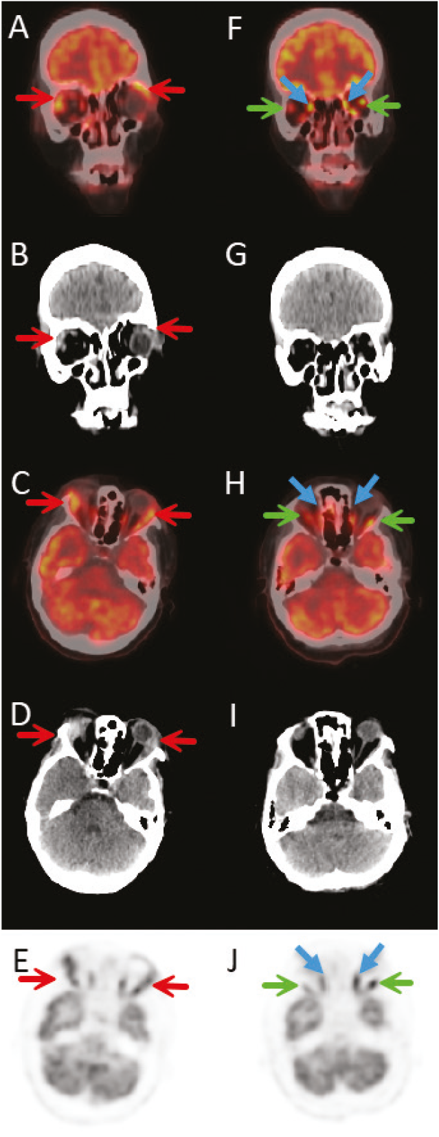
We report a case of IgG4-RD diagnosed by lacrimal gland biopsy. The patient presented with eyelid swelling and elevated inflammatory markers. 18F-FDG PET/CT was performed to assess systemic involvement, revealing increased FDG uptake and enlargement of both lacrimal glands, more pronounced on the right, without evidence of involvement of other organs.
The patient underwent corticosteroid and rituximab therapy. A follow-up 18F-FDG PET/CT conducted six months post-treatment demonstrated normalization of the lacrimal gland size and the absence of FDG uptake, indicating a favorable therapeutic response. To our knowledge, this is the first 18F-FDG PET/CT image of isolated lacrimal IgG4-RD demonstrating its complete response to treatment.
This case underscores the significance of 18F-FDG PET/CT in evaluating treatment response in IgG4-RD. Pre-treatment coronal fusion (A), coronal CT (B), axial fusion (C), axial CT (D), and axial PET (E) images demonstrate increased size and FDG uptake in the bilateral lacrimal glands, more prominent on the right (red arrows). Post-treatment coronal fusion (F), coronal CT (G), axial fusion (H), axial CT (I), and axial PET (J) images show normalization of lacrimal gland size and absence of pathological FDG uptake. Physiological uptake in lateral rectus (green arrows) and medial rectus (blue arrows) muscles can also be seen.
Footnotes
Ethics
Informed Consent: Informed consent was obtained from the patient for the use of their imaging data.
Authorship Contributions
Surgical and Medical Practices: A.K., S.A., Concept: A.K., S.A., O.E.Ş., L.U.B., S.S., K.S., H.B.S., Design: A.K., S.A., Data Collection or Processing: A.K., S.A., Analysis or Interpretation: A.K., S.A., O.E.Ş., L.U.B., S.S., K.S., H.B.S., Literature Search: A.K., Writing: A.K.
Conflict of Interest: No conflicts of interest were declared by the authors.
Financial Disclosure: The authors declare that this study has received no financial support.
References
Ren H, Mori N, Sato S, Mugikura S, Masamune A, Takase K. American College of Rheumatology and the European League Against Rheumatism classification criteria for IgG4-related disease: an update for radiologists. Jpn J Radiol. 2022;40:876-893.
Hao FY, Yang FX, Bian HY, Zhao X. Immunoglobulin G4-related lymph node disease with an orbital mass mimicking castleman disease: a case report. World J Clin Cases. 2021;9:10999-11006.
Perugino CA, Stone JH. IgG4-related disease: an update on pathophysiology and implications for clinical care. Nat Rev Rheumatol. 2020;16:702-714.
Sekiguchi H, Horie R, Kanai M, Suzuki R, Yi ES, Ryu JH. IgG4-related disease: retrospective analysis of one hundred sixty-six patients. Arthritis Rheumatol. 2016;68:2290-2299.
Kuroda N, Nao T, Fukuhara H, Karashima T, Inoue K, Taniguchi Y, Takeuchi M, Zen Y, Sato Y, Notohara K, Yoshino T. IgG4-related renal disease: clinical and pathological characteristics. Int J Clin Exp Pathol. 2014;7:6379-6385.
Wallace ZS, Naden RP, Chari S, Choi H, Della-Torre E, Dicaire JF, Hart PA, Inoue D, Kawano M, Khosroshahi A, Kubota K, Lanzillotta M, Okazaki K, Perugino CA, Sharma A, Saeki T, Sekiguchi H, Schleinitz N, Stone JR, Takahashi N, Umehara H, Webster G, Zen Y, Stone JH; American College of Rheumatology/European League Against Rheumatism IgG4-Related Disease Classification Criteria Working Group. The 2019 American College of Rheumatology/European League Against Rheumatism classification criteria for IgG4-related disease. Arthritis Rheumatol. 2020;72:7-19.
Tang CYL, Chua WM, Cheng LTJ, Fong W, Zaheer S, Lam WW. 18 F-FDG PET/CT manifestations of IgG4-related disease. Br J Radiol. 2021;94:20210105.
Zhang J, Chen H, Ma Y, Xiao Y, Niu N, Lin W, Wang X, Liang Z, Zhang F, Li F, Zhang W, Zhu Z. Characterizing IgG4-related disease with 18 F-FDG PET/CT: a prospective cohort study. Eur J Nucl Med Mol Imaging. 2014;41:1624-1634.
Bai Z, Zhou T, Yu Z, Chen Y, Dong L. Clinical value of 18 F-FDG PET/CT in IgG4-related disease. Ann Nucl Med. 2022;36:651-660.
Mitamura K, Arai-Okuda H, Yamamoto Y, Norikane T, Takami Y, Fujimoto K, Wakiya R, Ozaki H, Dobashi H, Nishiyama Y. Disease activity and response to therapy monitored by [ 18 F]FDG PET/CT using volume-based indices in IgG4-related disease. EJNMMI Res. 2020;10:153.
Ebbo M, Grados A, Guedj E, Gobert D, Colavolpe C, Zaidan M, Masseau A, Bernard F, Berthelot JM, Morel N, Lifermann F, Palat S, Haroche J, Mariette X, Godeau B, Bernit E, Costedoat-Chalumeau N, Papo T, Hamidou M, Harlé JR, Schleinitz N. Usefulness of 2-[ 18 F]-fluoro-2-deoxy-D-glucose-positron emission tomography/computed tomography for staging and evaluation of treatment response in IgG4-related disease: a retrospective multicenter study. Arthritis Care Res (Hoboken). 2014;66:86-96.
Fu Z, Chen G, Liu M, Li Z, Li Q. 18 F-FDG PET/CT findings in a patient with IgG4-related sclerosing mesenteritis. Clin Nucl Med. 2018;43:294-295.
Elsobky S, Dattani V, Wagner T, Mandumula S, Tavare A. FDG PET/CT in sacroiliac IgG4 disease. Clin Nucl Med. 2021;46:e548-e550.

