Incidentally Detected High-Grade Follicular Derived Non-Anaplastic Thyroid Carcinoma on 68Ga-PSMA PET-CT
PDF

Interesting Image

Incidentally Detected High-Grade Follicular Derived Non-Anaplastic Thyroid Carcinoma on 68Ga-PSMA PET-CT

Mol Imaging Radionucl Ther 2025;34(2):159-161 DOI: 10.4274/mirt.galenos.2025.46693
, , , ,
PDF
Permissions

Abstract

68Ga-prostate-specific membrane antigen (PSMA)-11 positron emission tomography/computed tomography (PET/CT) is currently routinely used for the evaluation of prostate cancer. However, due to its whole-body imaging capability, it can incidentally identify pathologies beyond prostate cancer. Herein, we describe a case of a 67-year-old man who was recently diagnosed with prostate carcinoma (PCa). The patient had a high PSMA-avid thyroid carcinoma detected incidentally during the initial staging of PCa with 68Ga-PSMA-11 PET-CT.

Keywords: Thyroid carcinoma, incidental, prostate-specific membrane antigen, neovasculature

,
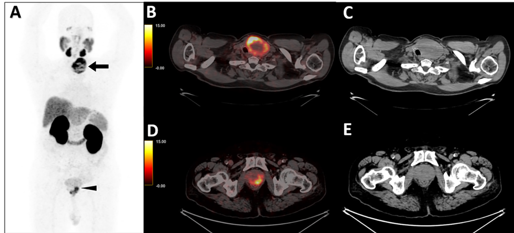
Figure 1
Figure 1. A 67-year-old gentleman with recently diagnosed prostate carcinoma (PCa) (Gleason’s score: 5+4=9) and serum prostate-specific antigen level 106 ng/mL underwent 68Ga-prostate-specific membrane antigen (PSMA)-11 positron emission tomography/computed tomography for staging. A) maximum intensity projection image showing a large focus of abnormal tracer uptake in the neck (arrow) and focal tracer uptake in the pelvis (arrowhead). B-C) The uptake in the neck localized to a large heterogeneous density mass with central necrosis and foci of calcification, replacing almost the entire left lobe of the thyroid and displacing the trachea toward the right measuring 6.8x5.4x7.1 cm with SUVmax 27.0 B-C). Pelvic PSMA uptake localized to an ill-defined lesion involving the bilateral peripheral zone of the prostate suggestive of known primary malignancy. D-E) The patient underwent ultrasonography (USG) neck which revealed a large thyroid mass on the thyroid imaging reporting and data system IV. Subsequently, the patient underwent USG-guided fine needle aspiration, which revealed a follicular neoplasm (Bethesda category-IV). Subsequently, the patient underwent total thyroidectomy, and histopathology revealed a high-grade follicular-derived non-anaplastic thyroid carcinoma.
View larger Download
Figure 2
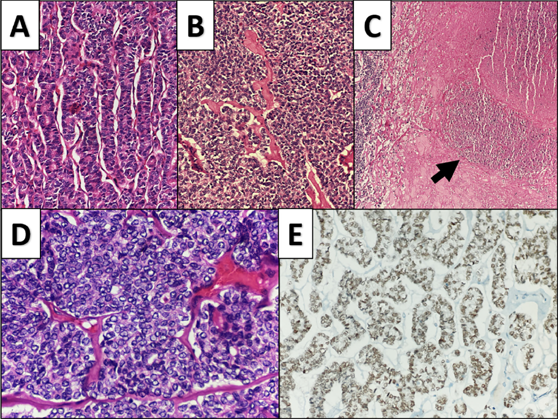
Figure 2. Histopathological images of thyroid tissue showing follicular cells arranged in a; A) trabecular growth pattern; B) solid, nested growth pattern H&E stain, 200x. C) Foci of necrosis are noted within the tumor. H&E stain, 40x. D) Nuclear features of papillary carcinoma of the thyroid, e.g., nuclear crowding, clearing, orphan annie nucleus and grooves H&E stain, 400x. E) Tumor cells showing immunoreactivity for Thyroid transcription factor-1, 200x. The features are suggestive of high-grade follicular-derived non-anaplastic thyroid carcinoma. PSMA is a type II transmembrane glycoprotein. This is overexpressed in prostate carcinoma (PCa) cells, more so in hormone-refractory PCa. PSMA overexpression is not restricted to the prostate and is also expressed in other normal tissues like salivary glands and duodenum. Further, it is overexpressed in endothelial cells of tumor neovasculature in different cancers of kidney, colon, melanoma, breast, etc. and also in benign diseases like endochondroma (,,,). PSMA is proven to be overexpressed on the endothelial cells of the neovasculature where it expedites endothelial cell sprouting and invasion to cleave the extracellular matrix by lytic proteases (). It is necessary to be aware of these pitfalls and to evaluate the incidental PSMA uptake apart from its physiological distribution, as in the present case in which abnormal tracer accumulation in the thyroid helped in the diagnosis of thyroid malignancy.
View larger Download

Footnotes

Ethics

Informed Consent: Informed consent was taken.

Authorship Contributions

Surgical and Medical Practices: P.S., S.M., A.K.A., G.K.P., K.A., Concept: P.S., S.M., A.K.A., Design: P.S., K.A., Data Collection or Processing: P.S., S.M., A.K.A., Analysis or Interpretation: P.S., S.M., A.K.A., K.A., Literature Search: P.S., S.M., Writing: P.S., S.M., A.K.A., G.K.P., K.A.

Conflict of Interest: No conflicts of interest were declared by the authors.

Financial Disclosure: The authors declare that this study has received no financial support.

References

1

Hofman MS, Hicks RJ, Maurer T, Eiber M. Prostate-specific membrane antigen PET: clinical utility in prostate cancer, normal patterns, pearls, and pitfalls. Radiographics. 2018; 38:200-217.

2

de Galiza Barbosa F, Queiroz MA, Nunes RF, Costa LB, Zaniboni EC, Marin JFG, Cerri GG, Buchpiguel CA. Nonprostatic diseases on PSMA PET imaging: a spectrum of benign and malignant findings. Cancer Imaging. 2020; 20:23.

3

Singh P, Kandula KK, Patel RK, Parida GK, Agrawal K. Incidental 68 Ga-PSMA uptake in the benign enchondroma-a rare finding on 68 Ga-PSMA PET/CT. Clin Nucl Med. 2024; 49:e525-e527.

4

Lauri C, Chiurchioni L, Russo VM, Zannini L, Signore A. PSMA expression in solid tumors beyond the prostate gland: ready for theranostic applications? J Clin Med. 2022; 11:6590.

5

Van de Wiele C, Sathekge M, de Spiegeleer B, De Jonghe PJ, Debruyne PR, Borms M, Beels L, Maes A. PSMA expression on neovasculature of solid tumors. Histol Histopathol. 2020; 35:919-927.