
Hepatic Vascular Shunts Mimicking Malignant Lesions on 18F-FDG PET/CT Imaging: Interpretation Pitfall in the Background of Cirrhotic Liver
PDF

Interesting Image

Hepatic Vascular Shunts Mimicking Malignant Lesions on 18F-FDG PET/CT Imaging: Interpretation Pitfall in the Background of Cirrhotic Liver

Mol Imaging Radionucl Ther 2026;35(1):61-63 DOI: 10.4274/mirt.galenos.2025.45712
, , , ,
PDF
Permissions

Abstract

A 44-year-old female presented with recent history of ischemic strokes and hypercoagulability and chronic alcoholic cirrhosis. 18F-fluorodeoxyglucose (FDG) positron emission tomography (PET)/computed tomography was performed during the workup for vasculitis. PET features of vasculitis were not identified, but liver showed multiple hepatic FDG avid foci, concerning for malignant or metastatic disease in the background of cirrhosis of liver. To characterize these lesions, liver magnetic resonance imaging was subsequently performed which revealed hepatic vascular shunts corresponding to the sites of the FDG avid foci. This case highlights potential interpretation n pitfall arising due to FDG avidity in the vascular shunts resembling malignant or metastatic lesions.

Keywords: 18F-FDG PET/CT, hepatic vascular shunt, liver cirrhosis
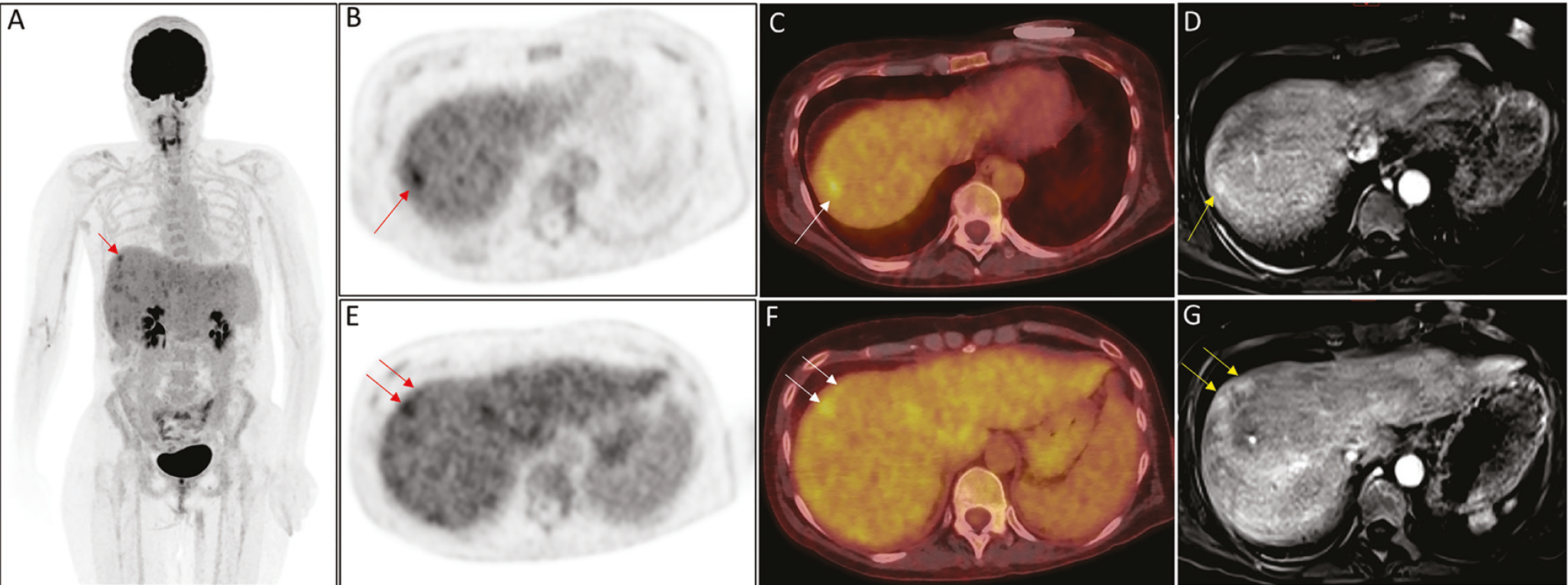
Figure 1
Figure 1. A 44-year-old woman presented with a history of recurrent ischemic strokes, alcoholic cirrhosis, hypercoagulability, and Raynaud’s phenomenon. A diagnostic angiogram showed multifocal narrowing of the bilateral middle cerebral arteries and their branches. Her serologic workup was remarkable for positive antinuclear antibodies, and anti-ribonucleoprotein antibodies. She also had positive cell based and immunofluorescence assays for cerebrospinal fluid glial fibrillary acidic protein. A contrast enhanced computed tomography (CECT) of chest, abdomen and pelvis was obtained which revealed lobulated liver with diffuse heterogeneous enhancement and no hepatic tumors; no significant findings related to vasculitis were noted. Subsequently, a whole body 18F-fluorodeoxyglucose positron emission tomography/computed tomography (18F-FDG PET/CT) was performed (,). The maximum intensity projection image (A) shows heterogeneous FDG distribution in the liver with few intensely FDG avid foci. Transaxial PET (B, E), and fused PET/CT (C, F) images shown here demonstrate the intensely FDG avid foci in the segments 7 and 8 of the liver (arrows), in the background of heterogeneous hepatic FDG uptake. No vasculitis pattern or other FDG avid abnormal foci were noted elsewhere. Given the history of cirrhosis, hypercoagulability and the clinical presentation, these FDG avid foci raised a concern for malignant or metastatic liver disease. Magnetic resonance imaging (MRI) the liver was then performed. Axial VIBE MRI sequences shown here (D, G) revealed subcapsular enhancing foci in the venous phase at the sites corresponding to FDG avid foci in hepatic segments 7 and 8, suggestive of vascular shunts. Furthermore, no arterially enhancing hepatic foci were seen. Intrahepatic vascular shunts can arise secondary to other various etiologies and hepatocellular carcinoma, hemangiomas, cirrhosis, trauma, congenital, Budd Chiari syndrome are among the common causes (). Cirrhosis is a known risk factor for developing hepatocellular carcinoma. In the presented case, both CECT and MRI revealed hepatic parenchymal changes and lobulated liver consistent with the history of alcoholic cirrhosis, but neither of these imaging modalities revealed hepatic tumors or metastatic lesions, nor any venous thrombosis was identified. As noted in this case, FDG avid foci in non-malignant conditions such as vascular shunts can resemble malignant or metastatic tumors, and it can be challenging to lean towards a diagnosis without further imaging (,). Dedicated MRI of the liver is often helpful in accurate characterization of such lesions (,,).This case highlights interpretation pitfall arising due to hepatic vascular shunt resembling malignant disease on 18F-FDG PET/CT in the background of cirrhotic liver, where malignancy is otherwise an important differential and utilizing multimodality imaging is advantageous in ruling out malignant disease.
View larger Download

Footnotes

Ethics

Informed Consent: Requirement for informed consent was waived by University of Iowa’s Human Subject Office.

Authorship Contributions

Concept: P.M., M.G., A.S., Design: A.S., Data Collection or Processing: S.J., A.S.E., Analysis or Interpretation: P.M., M.G., A.S., Literature Search: S.J., A.S.E., Writing: S.J., A.S.E.

Conflict of Interest: No conflicts of interest were declared by the authors.

Financial Disclosure: The authors declare that this study has received no financial support.

References

1

Lebech AM, Gaardsting A, Loft A, Graff J, Markova E, Bertelsen AK, Madsen JL, Andersen KF, Benzon EV, Helms M, Mathiesen LR, David KP, Kronborg G, Kjaer A. Whole-body 18F-FDG PET/CT is superior to CT as first-line diagnostic imaging in patients referred with serious nonspecific symptoms or signs of cancer: a randomized prospective study of 200 patients. J Nucl Med. 2017;58:1058-1064.

2

Slart RHJA; Writing group; Reviewer group; Members of EANM cardiovascular; members of EANM Infection & Inflammation; Members of Committees, SNMMI Cardiovascular; Members of Council, PET Interest Group; Members of ASNC; EANM Committee Coordinator. FDG-PET/CT(A) imaging in large vessel vasculitis and polymyalgia rheumatica: joint procedural recommendation of the EANM, SNMMI, and the PET Interest Group (PIG), and endorsed by the ASNC. Eur J Nucl Med Mol Imaging. 2018;45:1250-1269.

3

Wang Q, Koniaris LG, Milgrom DP, Patel A, Hu M, Cui E, Deng Y, Akisik F. CT and MRI imaging and interpretation of hepatic arterioportal shunts. Transl Gastroenterol Hepatol. 2019;4:34.

4

Ozaki K, Harada K, Terayama N, Kosaka N, Kimura H, Gabata T. FDG-PET/CT imaging findings of hepatic tumors and tumor-like lesions based on molecular background. Jpn J Radiol. 2020;38:697-718.

5

Tan GJ, Berlangieri SU, Lee ST, Scott AM. FDG PET/CT in the liver: lesions mimicking malignancies. Abdom Imaging. 2014;39:187-195.

6

Coenegrachts K. Magnetic resonance imaging of the liver: new imaging strategies for evaluating focal liver lesions. World J Radiol. 2009;1:72-85.

7

Bashir U, Shah S, Jeph S, O’Keeffe M, Khosa F. Magnetic resonance (MR) imaging of vascular malformations. Pol J Radiol. 2017;82:731-741.

8

Flors L, Leiva-Salinas C, Maged IM, Norton PT, Matsumoto AH, Angle JF, Hugo Bonatti M, Park AW, Ahmad EA, Bozlar U, Housseini AM, Huerta TE, Hagspiel KD. MR imaging of soft-tissue vascular malformations: diagnosis, classification, and therapy follow-up. Radiographics. 2011;31:1321-1340.

