

Abstract
Tuberculosis (TB) remains a significant public health problem in developing countries, but its diagnosis can be challenging as it may mimic malignancy or other granulomatous diseases. Although pulmonary TB is the most common form, TB can spread hematogenously, lymphatically, or by direct extension to any tissue or organ. We report a 62-year-old female who underwent 18F-fluorodeoxyglucose positron emission tomography/computed tomography (18F-FDG PET/CT) for malignancy evaluation, was diagnosed with extrapulmonary TB, and had her treatment response assessed with 18F-FDG PET/CT.
Keywords: Tuberculosis, malignancy, FDG PET, extrapulmonary, PET/CT
,
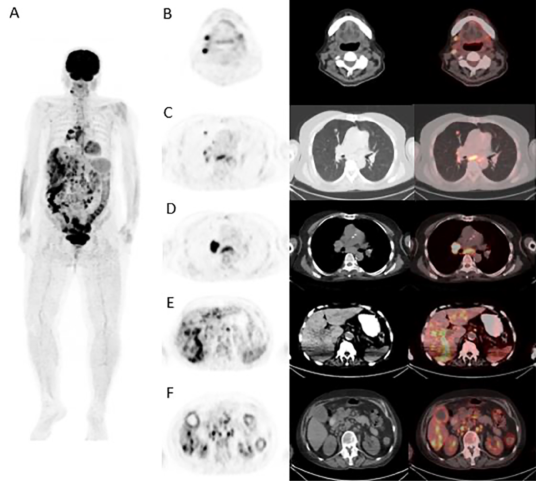
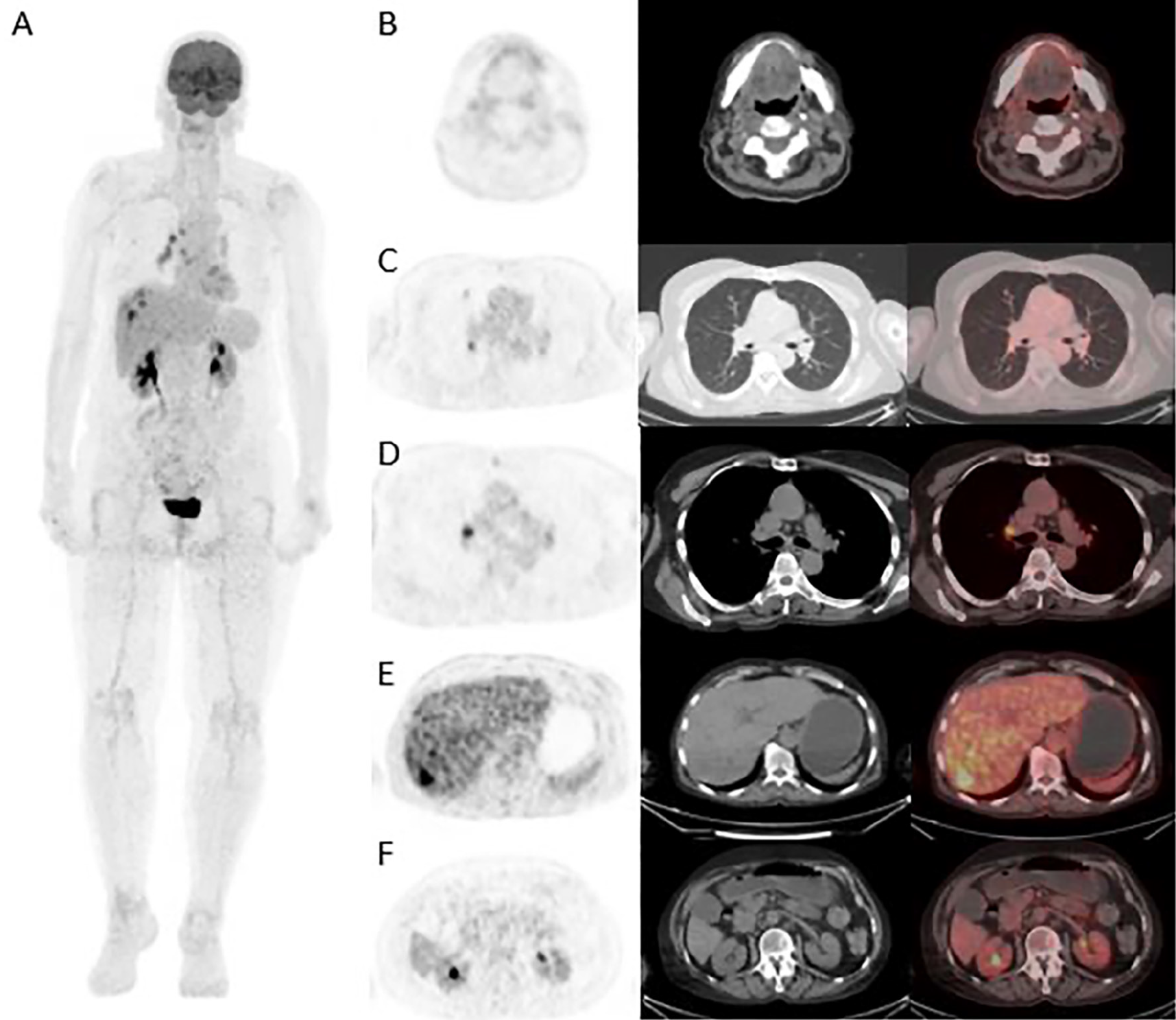
Although pulmonary TB is the most common form of the disease, TB can spread hematogenously, lymphatically, or by direct extension to virtually any tissue or organ. Hepatobiliary TB is a rare extrapulmonary manifestation that can mimic malignancy and may lead to diagnostic delays due to its nonspecific symptoms. TB should be considered in the differential diagnosis of patients, particularly immunocompromised individuals, presenting with multiple hypermetabolic hepatic lesions (,). 18F-FDG PET/CT is a valuable modality for detecting tuberculous lymphadenitis and serves as a complementary tool in the diagnosis of extrapulmonary TB. Owing to the intense FDG uptake by tuberculous granulomas, it is highly effective in identifying tuberculous lymphadenitis. Additionally, maximum standard uptake values provides information on the degree of inflammatory activity even in the absence of bacteriological confirmation. 18F-FDG PET/CT can detect lesions that may be overlooked on morphological imaging, assist in selecting optimal biopsy sites, and differentiate active from inactive disease by evaluating early treatment response. However, 18F-FDG PET/CT cannot reliably distinguish tuberculous lymphadenitis from lymphoma, sarcoidosis, or metastatic lymphadenopathy. In countries where TB is endemic, tuberculous lymphadenitis should be considered in the differential diagnosis of patients with enlarged lymph nodes, and lymph node biopsy plays a crucial role in establishing the diagnosis in light of 18F-FDG PET/CT findings (-).
Footnotes
Ethics
Informed Consent: Informed consent was obtained from the patient for publication.
Authorship Contributions
Concept: R.L.U.B., H.B.S., Design: R.L.U.B., H.B.S., Data Collection or Processing: C.G., Literature Search: C.G., Writing: C.G., R.L.U.B., H.B.S.
Conflict of Interest: No conflicts of interest were declared by the authors.
Financial Disclosure: The authors declare that this study has received no financial support.
References
Wang X, Shi X, Yi C, Chen Z, Zhang B, Zhang X. Hepatic tuberculosis mimics metastasis revealed by 18F-FDG PET/CT. Clin Nucl Med. 2014;39:e325-e327.
Di Renzo C, Tabrizian P, Kozuch DE, Fiel MI, Schwartz ME. Abdominal tuberculosis mimicking cancer clinically and on fluorodeoxyglucose (FDG)-positron emission tomography (PET) imaging: a two-case series. Am J Case Rep. 2020;21:e918901.
Liao F, Huang Z, Xu R, Luo Z, Qi W, Fan B, Yu J. Analysis of misdiagnosis and 18F-FDG PET/CT findings of lymph node tuberculosis. J Xray Sci Technol. 2022;30:941-951.
Yu WY, Lu PX, Assadi M, Huang XL, Skrahin A, Rosenthal A, Gabrielian A, Tartakovsky M, Wáng YXJ. Updates on 18 F-FDG-PET/CT as a clinical tool for tuberculosis evaluation and therapeutic monitoring. Quant Imaging Med Surg. 2019;9:1132-1146.
Sathekge M, Maes A, D’Asseler Y, Vorster M, Gongxeka H, Van de Wiele C. Tuberculous lymphadenitis: FDG PET and CT findings in responsive and nonresponsive disease. Eur J Nucl Med Mol Imaging. 2012;39:1184-1190.

