

Abstract
Lung cancer is the leading cause of cancer-related deaths worldwide. While metastasis to distant organs is commonly described, renal metastasis remains rare and uncommon. In this paper, we present the case of a 53-year-old man with a history of left apical lung adenocarcinoma who underwent fluorodeoxyglucose (FDG) positron emission tomography/computed tomography (CT) for suspected recurrence. The scan revealed a recurrent pulmonary mass with metastatic spread, notably showing intense bilateral focal FDG uptake in the kidneys. A metastatic origin was suspected and subsequently confirmed by CT-guided renal biopsy.
Although rare and typically observed in advanced stages of the disease, this case underscores the importance of carefully evaluating focal FDG uptake in the renal cortex to ensure accurate staging and optimal management of oncologic patients.
Keywords: Fluorodesoxyglucose, positron emission tomography/CT, lung adenocarcinoma, renal metastasis
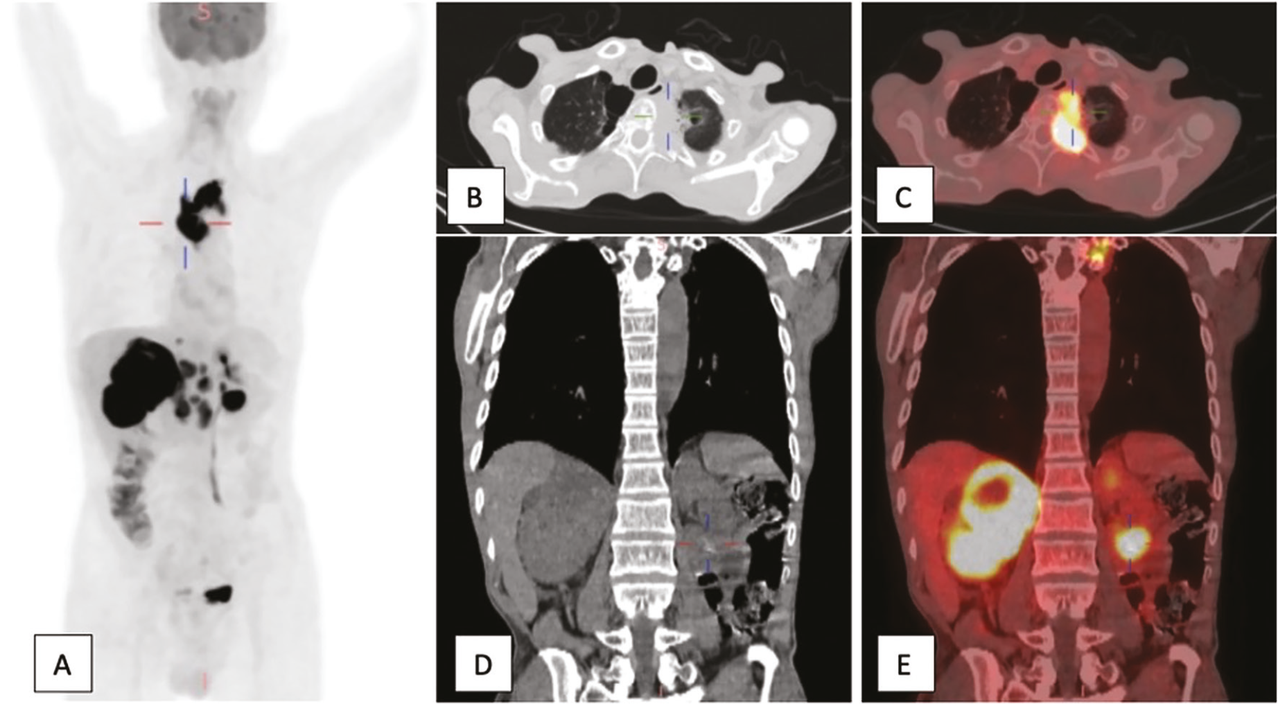
A 53-year-old man with a history of poorly differentiated adenocarcinoma of the left lung underwent whole-body 18F-FDG PET/CT for suspected recurrence. The PET scan, coupled with a low-dose CT scan (dose length product =1321 mGy:cm), was performed 60 minutes after the injection of 6.7 mCi of FDG. It revealed a hypermetabolic left upper-lobe lung mass [maximum standard uptake values (SUVmax) =21.5] with contiguous vertebral involvement (B, C) and multiple supradiaphragmatic and infradiaphragmatic lymphadenopathy. Notably, bilateral renal involvement was identified: a large, isodense, hypermetabolic mass infiltrating the right kidney and the adrenal gland (SUVmax =22) and a second hypodense lesion demonstrating intense focal FDG uptake (SUVmax =22.6) in the lower pole of the left renal cortex (D,E). A CT-guided renal biopsy was performed, and immunohistochemical analysis confirmed metastatic adenocarcinoma of pulmonary origin. This resulted in disease upstaging and initiation of systemic pemetrexed monotherapy. Renal metastases are an uncommon finding; historically, they have been diagnosed only post-mortem and typically originate from lung tumors (). They are often clinically silent, may be solitary, multifocal, unilateral, or bilateral, are difficult to distinguish from primary renal malignancies on conventional imaging, and are most often observed in advanced stages of the disease, generally in the context of widespread dissemination (). 18F-FDG PET/CT, commonly used in staging and restaging of lung cancer, plays a crucial role in identifying unusual metastatic patterns (); its sensitivity in the detection of renal lesions is, however, limited due to the physiological excretion of 18F-FDG through the urinary tract and depends on lesion size and location (). Although kidney metastases are rare, the few reported cases of kidney metastases highlight that renal FDG uptake can be overlooked or misinterpreted as tracer retention because of the high background activity (,). To date, there are no specific PET/CT findings that reliably distinguish renal metastases from primary renal tumors. In this case, a metastatic origin was strongly suspected due to bilateral, large, and intensely hypermetabolic renal lesions, their hypodense appearance on CT, and the known metastatic spread to other organs. Nevertheless, histological confirmation was essential to exclude synchronous primary renal tumors, thereby ensuring adequate staging of the disease and the implementation of appropriate treatment strategies.
Footnotes
Ethics
Informed Consent: Informed consent was obtained from the patient for publication.
Authorship Contributions
Surgical and Medical Practices: I.E.M., A.M., I.G., H.G., Concept: I.E.M., A.M., K.B., H.G., Design: I.E.M., K.B., Data Collection or Processing: I.E.M., A.M., Analysis or Interpretation: I.E.M., A.M., I.G., H.G., Literature Search: I.E.M., Writing: I.E.M., A.M.
Conflict of Interest: No conflicts of interest were declared by the authors.
Financial Disclosure: The authors declare that this study has received no financial support.
References
Lian H, Pan X, Hong B, Min J, Huang F. Metastases to the kidney from primary lung cancer: clinicopathological analysis of six cases in a single center. Diagn Pathol. 2023;18:60.
Zhou C, Urbauer DL, Fellman BM, Tamboli P, Zhang M, Matin SF, Wood CG, Karam JA. Metastases to the kidney: a comprehensive analysis of 151 patients from a tertiary referral centre. BJU Int. 2016;117:775-782.
Halaç M, Özhan M, Yilmaz Aksoy S, Vatankulu B, Aliyev A, Asa S, Atahan E, Sağer MS, Sönmezoğlu K. The role of FDG-PET/CT in detecting unsuspected and unknown distant metastasis in the initial staging of NSCLC. Turk J Med Sci. 2014;44:1029-1040.
Aras M, Dede F, Ones T, Inanır S, Erdil TY, Turoğlu HT. Is The value of FDG PET/CT in evaluating renal metastasis underestimated? A case report and review of the literature. Mol Imaging Radionucl Ther. 2013;22:109-112.
Zhang J, Dong A, Wang Y. FDG PET/CT in solitary isolated renal metastasis from squamous cell lung cancer. Clin Nucl Med. 2024;49:e50-e51.

