Analysis of Imaging Findings in a Patient with Squamous Cell Carcinoma of the External Auditory Canal Metastatic to the Dura with Trigeminal Nerve Involvement
PDF

Interesting Image

Analysis of Imaging Findings in a Patient with Squamous Cell Carcinoma of the External Auditory Canal Metastatic to the Dura with Trigeminal Nerve Involvement

Mol Imaging Radionucl Ther 2025;34(3):234-238 DOI: 10.4274/mirt.galenos.2025.26937
, , , ,
PDF
Permissions

Abstract

We report the case of a 56-year-old female recently diagnosed with well-differentiated squamous cell carcinoma of the external auditory canal. The patient underwent an 18F-fluorodeoxyglucose positron emission tomography/computed tomography for staging assessment. This examination revealed intense uptake in the right ear canals, tympanic cavity, eustachian canal, parapharyngeal area, and infratemporal fossa. Notably, we identified intracranial dural metastasis, which represents an uncommon site for metastatic spread in general.

Keywords: Squamous cell carcinoma of external auditory canal, dural metastasis, 18F-fluorodeoxyglucose positron emission tomography/computed tomography

, ,
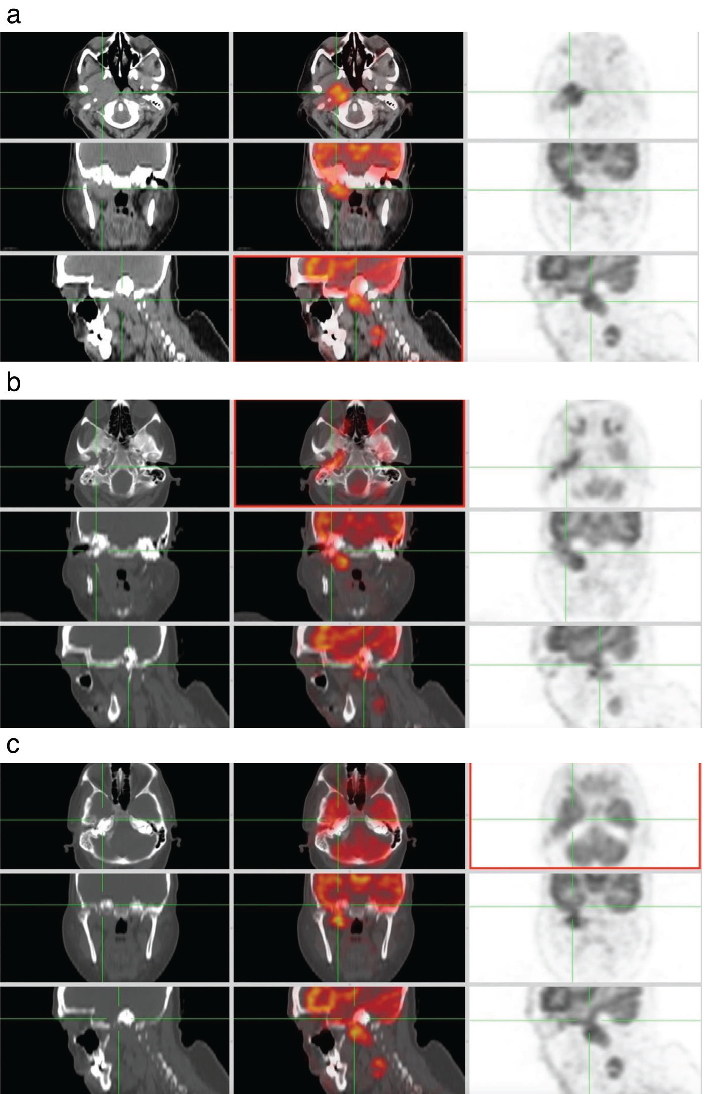
Figure 1
Figure 1. In a 56-year-old female patient diagnosed with well-differentiated squamous cell carcinoma of the external auditory canal, initial 18F-fluorodeoxyglucose positron emission tomography/computed tomography (18F-FDG PET/CT) images revealed a hypermetabolic mass standard uptake value maximum (SUVmax: 10.4, 40x30 mm in size) on the right side extending into the right parapharyngeal and masticator spaces and ipsilateral infratemporal fossa (Figure 1a). Increased 18F-FDG uptake was also observed extending linearly in the right tympanic cavity, ear canals (SUVmax: 8.0) and mastoid bone (SUVmax: 5.7) (Figure 1b). While intense physiological uptake by the brain presents a limitation in the evaluation of skull base lesions, skull base invasion of the tumor was clearly visible in the posteroinferior part of the temporal lobe in this case (SUVmax: 7.5, contralateral temporal lobe SUVmax: 5.7). Involvement of the right V3 nerve through the foramen ovale in Meckel’s cavity and intracranial spread were observed (Figure 1c). There was increased 18F-FDG uptake in a right cervical level 2, central hypometabolic peripheral hypermetabolic metastatic lymph node, 28x18 mm in size.
View larger Download
Figure 2
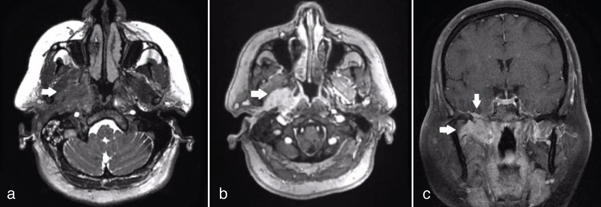
Figure 2. Magnetic resonance imaging examination revealed a 39x28 mm lesion, in the right parapharyngeal and masticator area, extending posterolaterally to the carotid sheath and posteriorly to the retropharyngeal area. The lesion appeared hypointense on T2-weighted images (Figure 2a) and exhibited dense heterogeneous contrast enhancement on post-contrast series (Figure 2b). The lesion involved the right pterygoid and masseter muscles, with intracranial extension via the right ınfratemporal fossa. Coronal T1-weighted contrast-enhanced imaging demonstrated skull base invasion through the foramen ovale, resulting in 6.5 mm dural thickening in the posteroinferior aspect of the temporal lobe (Figure 2c). Additionally, the lesion in the right tympanic cavity and external auditory canal (EAC) extended to the nasopharynx via the Eustachian tube, with intense T2 signal increases observed in these areas. Endoscopic nasopharyngeal examination revealed slight fullness in the right rosenmüller fossa. Otoscopic examination demonstrated diffuse maceration and bony erosion in the right EAC, as well as a thickened tympanic membrane. Punch biopsies were obtained from both the nasopharynx and the right EAC.
View larger Download
Figure 3
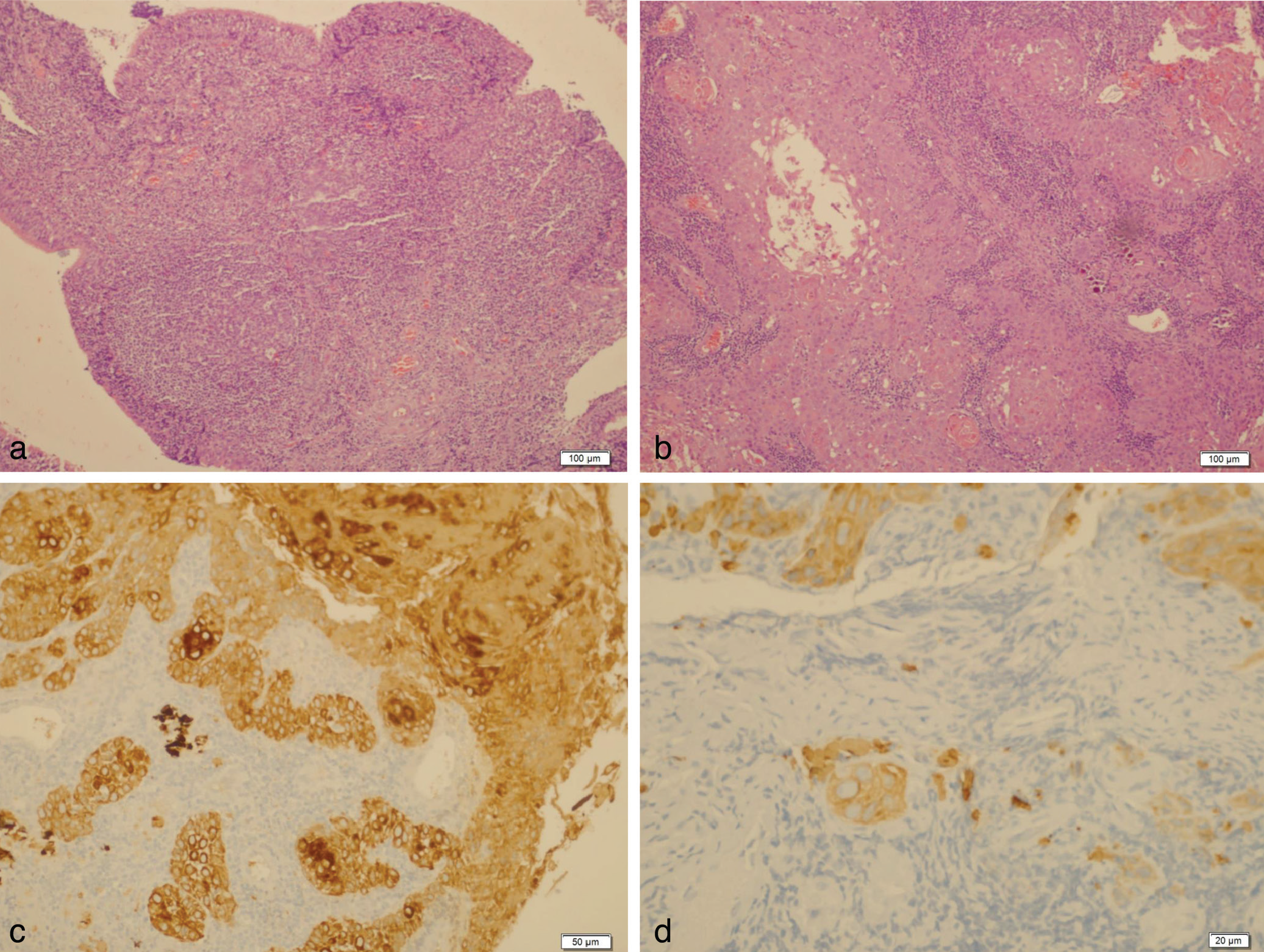
Figure 3. Lymphoid hyperplasia, focal squamous metaplasia/reactive cellular changes in the epithelium of the nasopharynx [Figure 3a,10x10 hematoxylin and eosin (HE)] and well-differentiated squamous cell carcinoma of the external auditory canal (EAC-SCC) were diagnosed (Figure 3b, 10x10 HE). Atypical squamous epithelial cells invading the subepithelial stroma of the EAC [(Figure 3c, 20x10 PanK immunohistochemistry (IHC)], (Figure 3d, 40x10 CK5/6 IHC). The patient was classified as T4N1M0 stage, and the tumor was deemed unresectable. Consequently, chemoradiotherapy was administered based on the decision of the multidisciplinary tumor board. Although primary malignant tumors of the EAC are rare (), T4 stage EAC-SCC exhibits a poor prognosis, with reported two-year survival rates for T4 stage EAC-SCC ranging from 0% to 7% (). Due to the continuity of the infratemporal fossa with the inferior parapharyngeal space, larger neoplasms can readily extend through the fat in the parapharyngeal space (). Perineural spread may occur through the foramina and fissures (). Cross-sectional imaging modalities such as 18F-FDG PET/CT and MRI play a crucial role in the accurate staging of EAC-SCC.
View larger Download

Footnotes

Ethics

Informed Consent: A consent form allowing the publication of imaging findings and images has been signed by the patient.

Authorship Contributions

Surgical and Medical Practices: G.S., İ.M.Ç., H.A., A.Ç., Concept: G.S., Design: G.S., Data Collection or Processing: G.S., İ.M.Ç., H.A., A.Ç., Analysis or Interpretation: G.S., İ.M.Ç., H.A., A.Ç., Literature Search: G.S., İ.M.Ç., H.A., Writing: G.S., İ.M.Ç., H.A., A.Ç., A.A.

Conflict of Interest: No conflicts of interest were declared by the authors.

Financial Disclosure: The authors declare that this study has received no financial support.

References

1

Ouaz K, Robier A, Lescanne E, Bobillier C, Morinière S, Bakhos D. Cancer of the external auditory canal. Eur Ann Otorhinolaryngol Head Neck Dis. 2013;130:175-182.

2

Moody SA, Hirsch BE, Myers EN. Squamous cell carcinoma of the external auditory canal: an evaluation of a staging system. Am J Otol. 2000;21:582-588.

3

Sah A, S TG, H CS. Imaging of the ınfratemporal fossa: a comprehensive pictorial essay. Indian J Surg Oncol. 2022;13:868-875.

4

Raut AA, Naphade PS, Chawla A. Imaging of skull base: pictorial essay. Indian J Radiol Imaging. 2012;22:305-316.