A Rare Case of Primary Squamous Cell Urachal Carcinoma Staged with 18F-FDG PET/CT
PDF

Interesting Image

A Rare Case of Primary Squamous Cell Urachal Carcinoma Staged with 18F-FDG PET/CT

Mol Imaging Radionucl Ther 2026;35(2):131-133 DOI: 10.4274/mirt.galenos.2025.21347
, , , , ,
PDF
Permissions

Abstract

Primary squamous cell urachal carcinoma is an exceedingly rare epithelial neoplasm and an aggressive malignancy, originating from the urachal remnants. In contrast to mucinous adenocarcinoma, the most common histological type, squamous cell urachal carcinoma demonstrates high 2-deoxy-2-18F-fluorodeoxyglucose (18F-FDG) uptake, making 18F-FDG positron emission tomography/computed tomography (PET/CT) a valuable tool for evaluating regional lymph node involvement and detecting distant metastases. Optimal surgical management, the need for systemic therapy, and the overall prognosis are largely determined by accurate clinical staging, which is mainly based on the widely recognized Sheldon and Mayo classifications. We present a rare case of primary squamous cell urachal carcinoma on 18F-FDG PET/CT in a young woman, with correlation to surgical and histopathological findings.

Keywords: Rare malignancy, primary urachal carcinoma, squamous cell carcinoma, 18F-FDG PET/CT

,
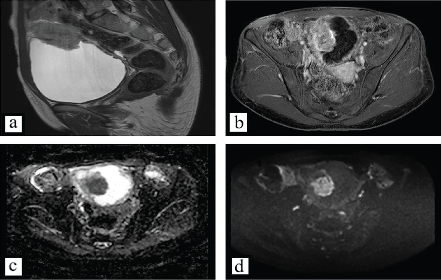
Figure 1
Figure 1. A 34-year-old woman presented with dysuria and recurrent urinary tract infections. Initial ultrasonography demonstrated a lesion in the bladder area. Subsequent magnetic resonance imaging (MRI) revealed a midline supravesical solid mass along the course of the urachal ligament and contiguous with the bladder dome, radiologically consistent with originating from urachal remnants. On MRI, the lesion appeared iso- to hypointense on T2-weighted sequences (Figure 1a), showed homogeneous post-gadolinium enhancement (Figure 1b), and showed restricted diffusion (Figures 1c,d ). Written informed consent was obtained from the patient for the publication of her clinical information and imaging data.
View larger Download
Figure 2
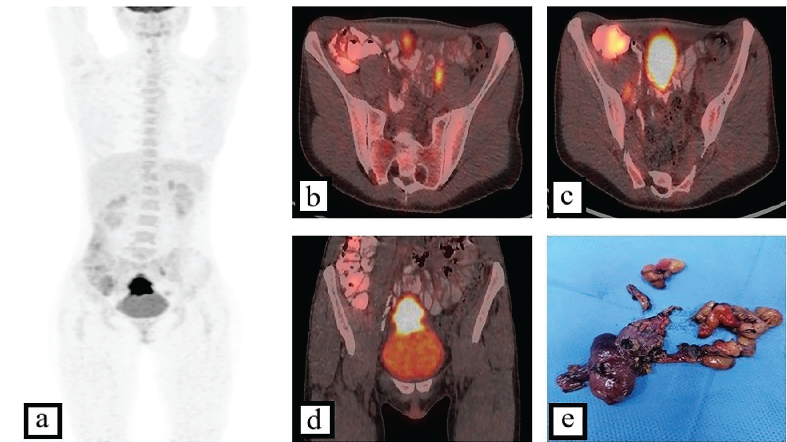
Figure 2. Cystoscopy confirmed a dome-based lesion, and transurethral biopsy specimens were obtained. Histopathological analysis revealed a squamous cell carcinoma. Primary urachal carcinoma is an uncommon malignancy arising from embryonic urachal remnants, accounting for less than 1% of all bladder cancers (). According to a recent meta-analysis of registry data published in 2024, adenocarcinoma represents the predominant histological type, comprising approximately 86% of cases. By contrast, squamous cell carcinoma is an exceedingly rare variant, representing only about 2% of urachal tumors, with no more than 30 cases documented in the literature to date (). The extreme rarity of this malignancy poses a significant therapeutic challenge, as its optimal management strategies remain poorly defined. The therapeutic approach and the overall prognosis are largely determined by clinical stage. Several staging systems have been proposed for urachal carcinoma, the most widely applied being those of Sheldon () and Mayo (). To facilitate accurate staging, the patient underwent whole-body positron emission tomography/computed tomography (PET/CT) with 2-deoxy-2-18F-fluorodeoxyglucose (18F-FDG). The maximum intensity projection image (Figure 2a) and the coronal fused PET/CT slice (Figure 2d) demonstrated a hypermetabolic solid soft-tissue mass extending along the urachal ligament to the bladder dome, with a maximum standardized uptake value (SUVmax) of 25.1. No evidence of distant metastatic disease was observed. However, axial fused PET/CT images of the pelvis revealed solitary 18F-FDG -avid lymph nodes (SUVmax up to 5.4) along the left (Figure 2b) and right (Figure 2c) external iliac vessels. Although the therapeutic value of lymphadenectomy in urachal carcinoma remains a matter of debate, the suspicion of regional nodal involvement warranted an aggressive surgical approach in this case. The subsequent surgical procedure consisted of partial cystectomy with en bloc resection of the urachal ligament and umbilicus, combined with extended pelvic lymph node dissection (Figure 2e). Histopathological examination confirmed a low-grade, pure squamous cell urachal carcinoma with negative surgical margins and no nodal metastases, corresponding to stage II according to the Mayo classification and stage IIIA according to the Sheldon classification. Because the disease was node-negative, adjuvant chemotherapy was not considered mandatory in accordance with the National Comprehensive Cancer Network guidelines (). Nevertheless, given the aggressive biological behavior of this malignancy and the marked glucose metabolism characteristic of this histological type, as demonstrated in the presented case, 18F-FDG PET/CT may serve as a valuable tool for surveillance, particularly when there is clinical suspicion of local recurrence or distant metastasis.
View larger Download

Footnotes

Ethics

Informed Consent: Written informed consent was obtained from the patient for the publication of her clinical information and imaging data.

Authorship Contributions

Surgical and Medical Practices: Y.G., A.S., S.A., N.H., Concept: Y.G., A.S., Design: Y.G., G.G., Data Collection or Processing: Y.G., G.G., N.H., Analysis or Interpretation: Y.G., Literature Search: Y.G., V.H., Writing: Y.G., V.H.

Conflict of Interest: No conflicts of interest were declared by the authors.

Financial Disclosure: The authors declare that this study has received no financial support.

References

1

Bruins HM, Visser O, Ploeg M, Hulsbergen-van de Kaa CA, Kiemeney LA, Witjes JA. The clinical epidemiology of urachal carcinoma: results of a large, population based study. J Urol. 2012;188:1102-1107.

2

Olah C, Kubik A, Mátrai P, Engh MA, Barna V, Hegyi P, Reis H, Nyirády P, Szarvas T. Estimation of the incidence of urachal cancer: a systematic review and meta-analysis of registry-based studies. Urol Oncol. 2024;42:221.e1-221.e7.

3

Sheldon CA, Clayman RV, Gonzalez R, Williams RD, Fraley EE. Malignant urachal lesions. J Urol. 1984;131:1-8.

4

Ashley RA, Inman BA, Sebo TJ, Leibovich BC, Blute ML, Kwon ED, Zincke H. Urachal carcinoma: clinicopathologic features and long-term outcomes of an aggressive malignancy. Cancer. 2006;107:712-720.

5

National Comprehensive Cancer Network. Bladder Cancer. Version 1.2025. NCCN Clinical Practice Guidelines in Oncology. Available at: https://www.nccn.org/professionals/physician_gls/pdf/bladder.pdf