

Abstract
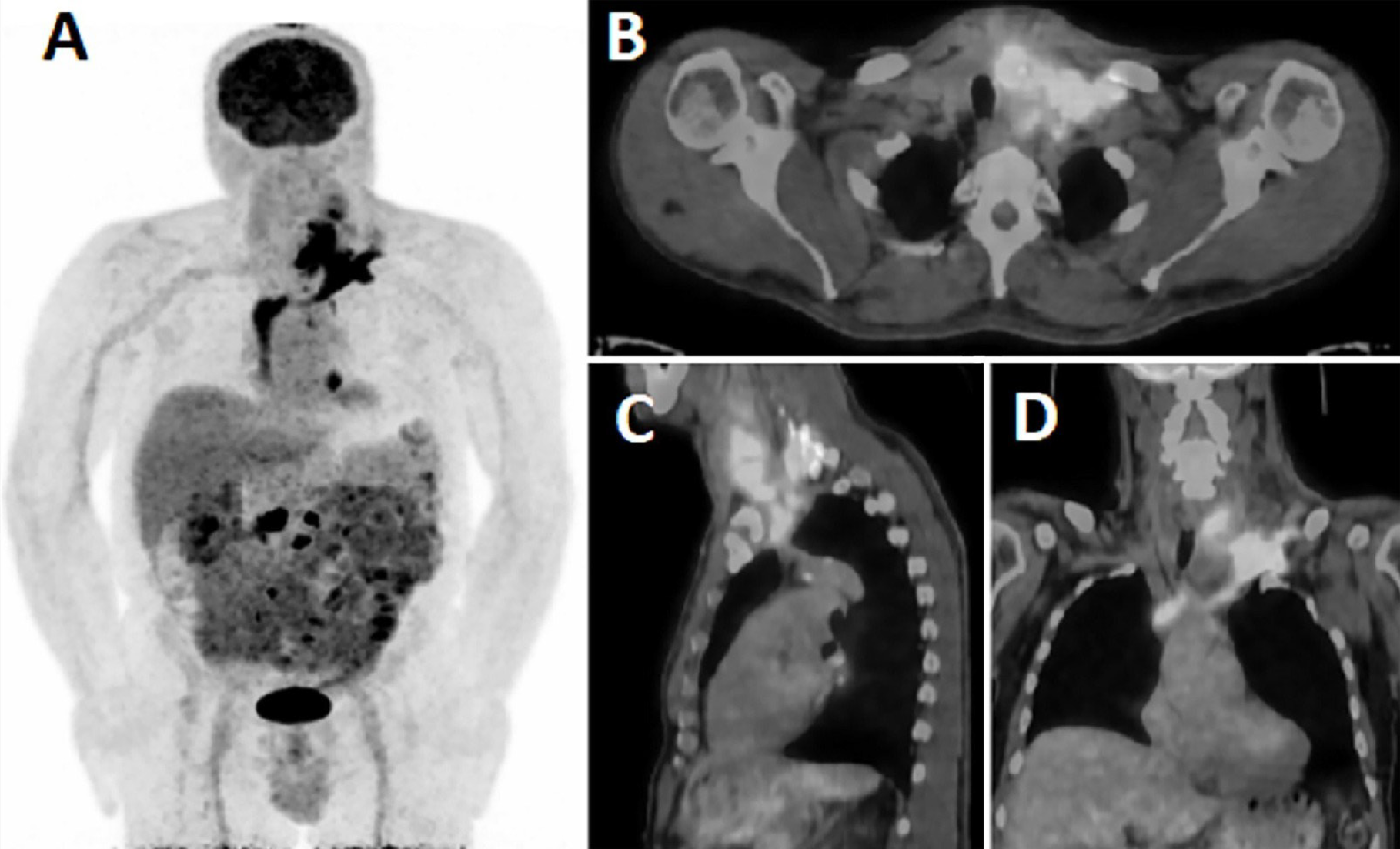
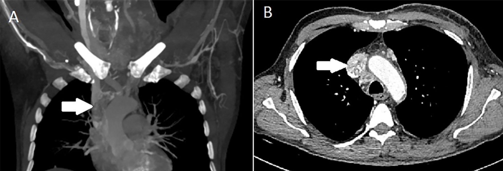
A 59-year-old man admitted to the hospital complained of left arm and neck pain. Magnetic resonance imaging was performed for possible cervical discopathy. It revealed that in the C7 vertebral body, lesions with surrounding bone marrow edema were observed, which were compatible with metastasis that caused cervical stenosis. Cervical stenosis surgery was performed, and the lesion was pathologically diagnosed as a metastasis of thyroid carcinoma. 18F-fluorodeoxyglucose positron emission tomography/computed tomography (18F-FDG PET/CT) scan was ordered for further evaluation. The PET/CT images showed an increased 18F-FDG uptake from the left internal jugular vein to the right atrium. It was considered a malignant thrombus.
Keywords: Malignant thrombus, 18F-FDG PET/CT, papillary thyroid cancer
,
Footnotes
Ethics
Informed Consent: The patient provided written informed consent for the publication of his images.
Authorship Contributions
Surgical and Medical Practices: M.N.C.T, L.P, H.Ö, G.K.G, Concept: M.N.C.T, L.P, H.Ö, G.K.G, Design: M.N.C.T, L.P, H.Ö, G.K.G, Data Collection or Processing: M.N.C.T, L.P, H.Ö, G.K.G, Analysis or Interpretation: M.N.C.T, L.P, H.Ö, G.K.G, Literature Search: M.N.C.T, L.P, H.Ö, G.K.G, Writing: M.N.C.T, L.P, H.Ö, G.K.G.
Conflict of Interest: No conflicts of interest were declared by the authors.
Financial Disclosure: The authors declare that this study has received no financial support.
References
Fotis T, Konstantinou E, Mariolis-Sapsakos T, Mitsos A, Restos S, Katsenis K, Elefsiniotis I, Kapellakis G. Solitary internal jugular vein invasion by thyroid carcinoma: resection and reconstruction. J Vasc Nurs. 2009;27:46-47.
Leong JL, Yuen HW, LiVolsi VA, Loevner L, Narula N, Baloch Z, Weber RS. Insular carcinoma of the thyroid with jugular vein invasion. Head Neck. 2004;26:642-646.
Taib NA, Hisham AN. Follicular thyroid carcinoma with direct tumour extension into the great cervical veins and right atrium: is transcervical thrombectomy a safe option? Asian J Surg. 2007;30:216-219.
Varghese BT, Sebastian P. Internal jugular vein thrombus associated with thyroid carcinoma. Ann Otol Rhinol Laryngol. 2005;114:656.
Koike E, Yamashita H, Watanabe S, Yamashita H, Noguchi S. Brachiocephalic vein thrombus of papillary thyroid cancer: report of a case. Surg Today. 2002;32:59-62.
Strobel K, Steinert HC, Bhure U, Koma AY, Gassmann N, Stöckli SJ. Tumour thrombus in the superior vena cava from anaplastic carcinoma of the thyroid: FDG-PET/CT imaging findings. Eur J Nucl Med Mol Imaging. 2007;34:813.

