Triple Tumors Uncovered: Insights from 68Ga PSMA PET-CT
PDF

Interesting Image

Triple Tumors Uncovered: Insights from 68Ga PSMA PET-CT

Mol Imaging Radionucl Ther 2025;34(3):224-227 DOI: 10.4274/mirt.galenos.2025.15870
, , ,
PDF
Permissions

Abstract

Galium-68 prostate-specific membrane antigen positron emission tomography combined with computed tomography (68Ga PSMA PET-CT) is receptor specific imaging, which has increasingly been used in the staging and restaging of prostate carcinoma (PCa). PSMA is type II transmembrane glycoprotein expressed in cytosol of normal prostatic tissue with 100-1000-fold over expression in PCa. It is also expressed in the endothelium of tumor-associated neo vasculature of non-prostatic solid tumor such as transitional cell and renal cell carcinoma, hepatocellular, thyroid, and brain cancers. We hereby present a case where PSMA PET scan showed three tumors proved on follow up imaging.

Keywords: 68Ga PSMA PET-CT, prostate carcinoma, hepatocellular carcinoma, meningioma

,
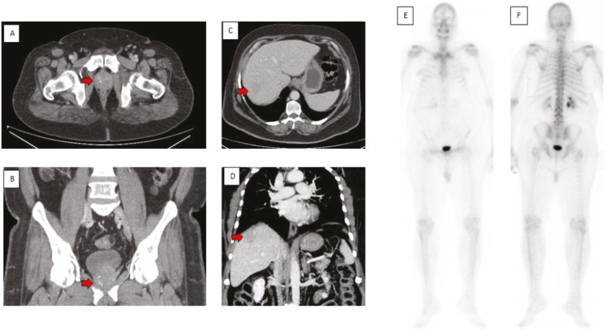
Figure 1
Figure 1. A 63-year-old gentleman, known as a diabetic and hypertensive, presented with pain in the right flank. He also had cirrhotic liver against background of hepatitis C virus infection. His ultrasound showed enlarged prostate gland, with prostate-specific antigen of 128 ng/mL  and histopathology showed prostatic adenocarcinoma, Gleason score 9/10, and World Health Organization group 5. His computed tomography (CT) chest, abdomen, and pelvis, in addition to the primary prostate carcinoma (PCa) (A, B), showed coarse liver parenchyma with hypodensity in segment VI/VII (C, D); differentials included metastasis or hepatocellular carcinoma. A 99mTc MDP bone scan was negative for osteoblastic metastasis (E, F). In view of a high Gleason score, the Galium-68 (68Ga) prostate-specific membrane antigen (PSMA) positron emission tomography (PET)/CT findings and the tumor board suggested a multiparametric magnetic resonance imaging. 68Ga PSMA PET/CT, due to its high sensitivity and specificity, is being increasingly used to stage PCa in intermediate - to high- and very high-risk patients when conventional imaging modalities are either negative for distant metastases or show fewer than 5 distant lesions (,). PSMA is a type II transmembrane glycoprotein with high expression in PCa and variable expression in neovasculature of some other tumors, such as hepatocellular, lung, thyroid, and renal cell carcinomas (-). This allows its potential role in the diagnosis and management of such tumors, which are typically non-avid on 18F-fluorodeoxyglucose PET/CT (18F-FDG) PET/CT which till now is the workhorse in the world of PET/CT (,).
View larger Download
Figure 2
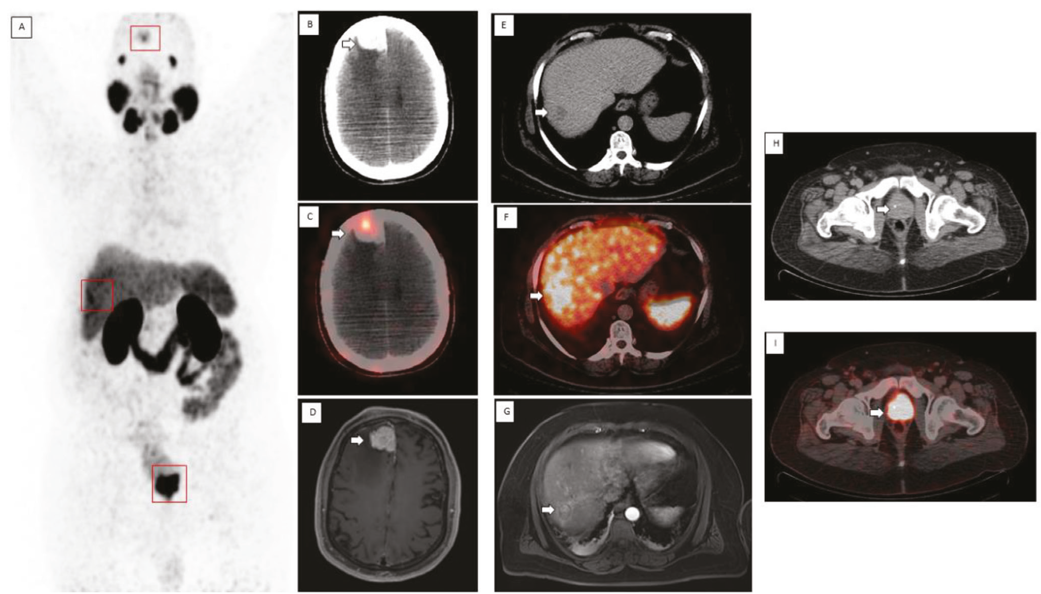
Figure 2. Maximum intensity projection image on Galium-68 prostate-specific membrane antigen positron emission tomography combined with computed tomography (68Ga PSMA PET/CT) (A) showed avid lesions in the brain, liver and pelvic region (red boxes). There was an enlarged PSMA-avid prostate gland along with an avid enhancing lesion in the right frontal lobe (B, C). His brain magnetic resonance imaging (MRI) with contrast showed an extra-axial mass in the right frontal region adjacent to the falx, suggestive of meningioma (D). In addition, increased PSMA uptake was seen in the known hepatic lesions (E, F). He also had multiparametric MRI with contrast showing pseudocapsular enhancement and early arterial phase enhancement within this lesion, which was highly suggestive of hepatocellular carcinoma (HCC) (G). Also, the primary prostate tumor showed increased PSMA uptake (H-I). Therefore, he was staged as T3N0M0 for prostate cancer and started on androgen deprivation therapy. Hepatobiliary surgeons evaluated his hepatic lesion, which led to resection of segments VI and VII. Histopathology confirmed it to be well-differentiated HCC. After 1.5 years, he received 6000 cGy of radiation in 20 fractions to the prostate gland and seminal vesicles. A few months later, he had stereotactic radiosurgery of 2700 cGy in 3 fractions, to the right frontal lobe meningioma. In our patient, 68Ga PSMA PET/CT was of paramount importance because it not only helped with the correct initial staging of PCa, but also detected two other tumors, i.e., HCC and meningioma. Therefore, we should be aware of these pitfalls of 68Ga PSMA PET/CT and be careful in labeling all PSMA avid lesions metastatic because all that glitters is not gold ().
View larger Download

Footnotes

Ethics

Informed Consent: Exemption of informed consent was sought from the institutional review board (IRB) [Exemption number- EX-29-04-20-04] based on the following points: a) It is a retrospective study, and information in this study is currently in existence in the hospital information system. b) The information is recorded in such a manner that the subjects cannot be identified. c) There would be no contact with the patient.

Authorship Contributions

Surgical and Medical Practices: S.M.G., A.H., W.A., I.U.D., Concept: S.M.G., W.A., Design: S.M.G., A.H., Data Collection or Processing: S.M.G., Analysis or Interpretation: S.M.G., A.H., W.A., I.U.D., Literature Search: S.M.G., Writing: S.M.G., A.H., W.A., I.U.D.

Conflict of Interest: No conflicts of interest were declared by the authors.

Financial Disclosure: The authors declare that this study has received no financial support.

References

1

Jadvar H, Calais J, Fanti S, Feng F, Greene KL, Gulley JL, Hofman M, Koontz BF, Lin DW, Morris MJ, Rowe SP, Royce TJ, Salami S, Savir-Baruch B, Srinivas S, Hope TA. appropriate use criteria for prostate-specific membrane antigen PET imaging. J Nucl Med. 2022;63:59-68.

2

Jochumsen MR, Bouchelouche K. PSMA PET/CT for primary staging of prostate cancer - an updated overview. Semin Nucl Med. 2024;54:39-45.

3

Kopka K, Benešová M, Bařinka C, Haberkorn U, Babich J. Glu-ureido-based inhibitors of prostate-specific membrane antigen: lessons learned during the development of a novel class of low-molecular-weight theranostic radiotracers. J Nucl Med. 2017;58:17S-26S.

4

Lisney AR, Leitsmann C, Strauß A, Meller B, Bucerius JA, Sahlmann CO. The role of PSMA PET/CT in the primary diagnosis and follow-up of prostate cancer-a practical clinical review. Cancers (Basel). 2022;14:3638.

5

Hangaard L, Jochumsen MR, Vendelbo MH, Bouchelouche K. Metastases from colorectal cancer avid on 68Ga-PSMA PET/CT. Clin Nucl Med. 2017;42:532-533.

6

Tariq A, Pearce A, Rhee H, Kyle S, Raveenthiran S, Pelecanos A, Gan CL, Goh JC, Wong D, McBean R, Marsh P, Goodman S, Dunglison N, Esler R, Navaratnam A, Yaxley JW, Thomas P, Pattison DA, Roberts MJ. The role of prostate-specific membrane antigen positron emission tomography/computed tomography in primary staging of selected renal tumours: initial experience in a multicentre cohort. Eur Urol Focus. 2024;10:770-778.

7

de Galiza Barbosa F, Queiroz MA, Nunes RF, Costa LB, Zaniboni EC, Marin JFG, Cerri GG, Buchpiguel CA. Nonprostatic diseases on PSMA PET imaging: a spectrum of benign and malignant findings. Cancer Imaging. 2020;20:23.

8

Voter AF, Werner RA, Savas H, Gafita A, Ross AE, Gorin MA, Solnes LB, Pomper MG, Rowe SP, Sheikhbahaei S. A practical guide to the pearls and pitfalls of PSMA PET imaging. Semin Nucl Med. 2024;54:119-131.