Hydatic Cyst Infection of the Tibia Mimicking Chondrosarcoma on 18F-FDG PET/CT Scan
PDF

Interesting Image

Hydatic Cyst Infection of the Tibia Mimicking Chondrosarcoma on 18F-FDG PET/CT Scan

Mol Imaging Radionucl Ther 2026;35(2):127-130 DOI: 10.4274/mirt.galenos.2025.13540
, , , , ,
PDF
Permissions

Abstract

A hydatid cyst is a zoonotic infection caused by Echinococcus granulosus. The liver and lungs are the most affected organs. Although rare, osseous involvement by hydatid cyst disease can occur because the rigid bone trabeculae prevent formation of the characteristic adventitia. While fluorodeoxyglucose (FDG) positron emission tomography (PET) findings in hepatic and pulmonary hydatid cysts are well described, this case, to the best of our knowledge, represents the first report of bone hydatid disease located in an extremity evaluated by 18F-FDG PET/computed tomography.

Keywords: Hytadic cyst, bone diseases, infection, 18F-FDG PET/CT

, ,
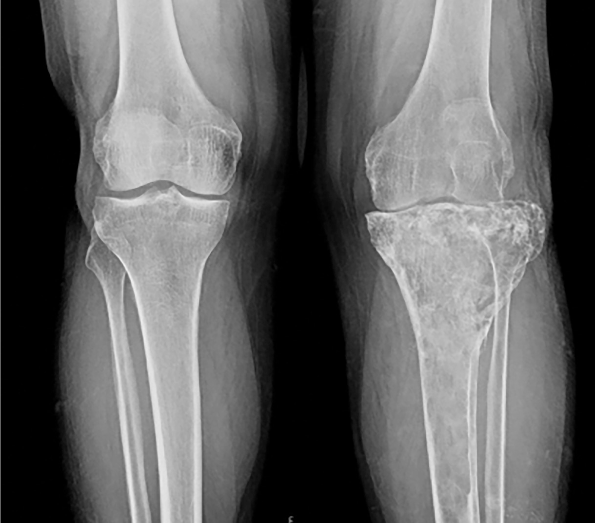
Figure 1
Figure 1. A 54-year-old male patient presented with progressively worsening pain and swelling localized to the left knee. Initial radiological evaluation using anteroposterior X-ray imaging demonstrated extensive lytic-destructive lesions predominantly involving the proximal metaphysis of the left tibia. Radiographic features included marked trabecular disruption, cortical thinning, and expansion of the medullary cavity. Given these aggressive radiological findings, a primary bone malignancy, particularly chondrosarcoma, was considered a preliminary diagnosis.
View larger Download
Figure 2
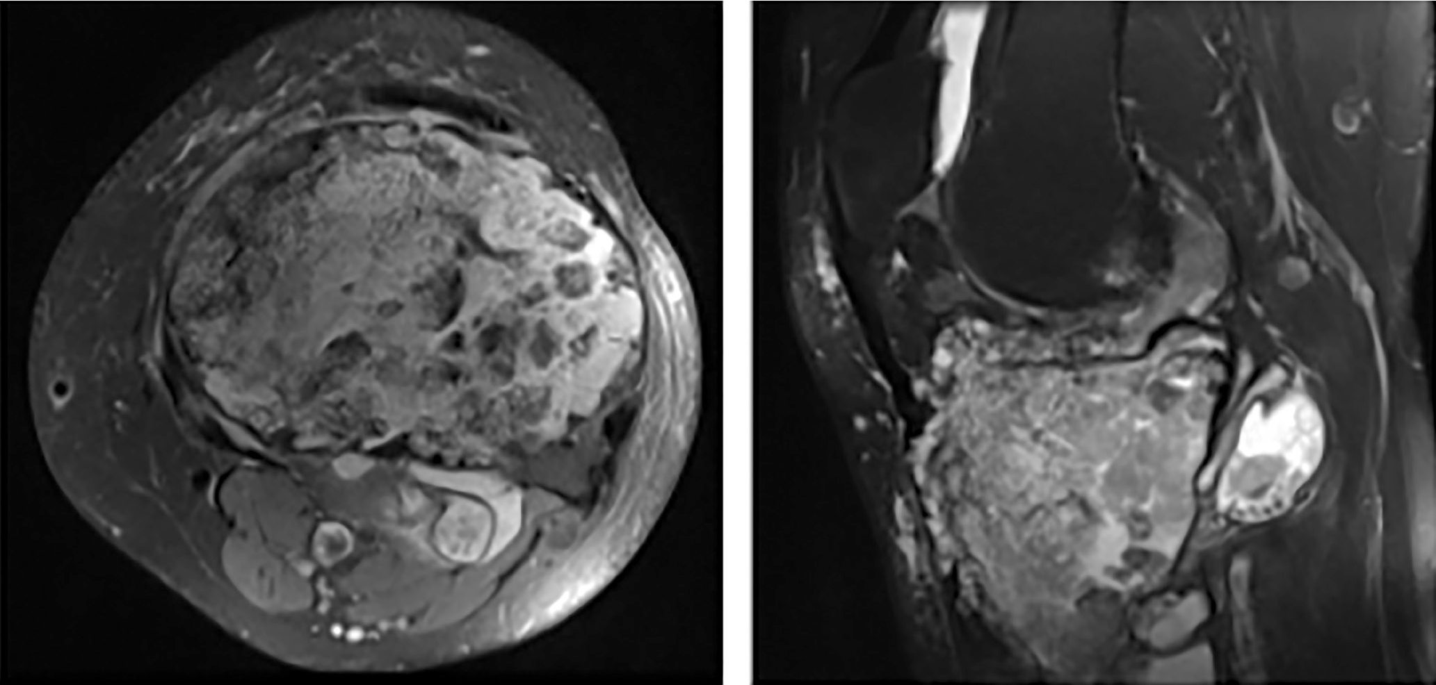
Figure 2. Subsequent magnetic resonance imaging (MRI) was conducted to further characterize the lesion. Axial (A) and sagittal (B) T2-weighted MRI sequences revealed a large, cystic lesion localized at the epiphyseal-metaphyseal junction of the proximal tibia. The lesion exhibited characteristic internal septations, focal chondroid-like calcifications, and prominent soft-tissue infiltration extending beyond the bone marrow cavity into surrounding musculature and adipose tissue. These radiological findings strongly supported the initial clinical suspicion of chondrosarcoma, necessitating further staging investigations.
View larger Download
Figure 3
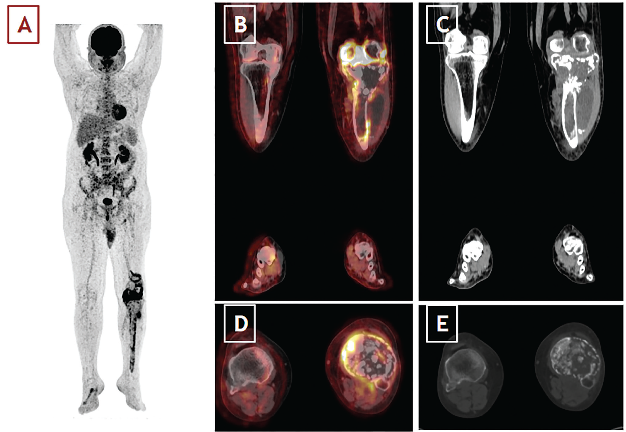
Figure 3. To assess systemic involvement and metastatic potential, an 18F-fluorodeoxyglucose positron emission tomography/computed tomography (18F-FDG PET/CT), scan was performed. Maximum-intensity projection imaging (A) identified intensely hypermetabolic foci (maximum standard uptake values: 27.3) in the proximal left tibia and distal femur. On coronal (B, C) and axial (D, E) fused PET/CT images, the lesions demonstrated marked cortical destruction with irregular margins and peripheral, heterogeneous FDG uptake. Within the bone marrow, cystic and lytic areas showed relatively low or absent tracer accumulation, suggestive of necrotic or fluid-filled components, whereas the surrounding soft-tissue extensions were markedly hypermetabolic and contained partially calcified densities. Mild periosteal reaction and adjacent soft-tissue edema were also evident, indicating biologic aggressiveness. Additionally, mild FDG uptake was noted in the left inguinal lymph nodes and was interpreted as reactive rather than metastatic. Because of persistent suspicion of malignancy on imaging, a tru-cut biopsy was performed. Histopathological analysis unexpectedly identified a solitary osseous hydatid cyst (caused by Echinococcus granulosus). No additional disease foci were detected upon further systemic evaluation with abdominal and thoracic CT imaging. The patient received antiparasitic therapy with albendazole, followed by successful surgical excision of the lesion. Although rare, hydatid cyst disease can also involve osseous structures because the rigidity of trabecular bone prevents formation of the characteristic adventitia 1-5. While FDG PET findings in hepatic 6 and pulmonary 7 hydatid cysts are well described, this case highlights the importance of considering osseous hydatid disease in the differential diagnosis of aggressive bone lesions, particularly in endemic regions.
View larger Download

Footnotes

Ethics

Informed Consent: Informed consent was obtained from the patient for publication of this case and accompanying images.

Authorship Contributions

Surgical and Medical Practices: H.K., B.D., Design: B.K., Data Collection or Processing: F.A., İ.T., Analysis or Interpretation: İ.T., Literature Search: F.A., Z.Ö., Writing: F.A., Z.Ö.

Conflict of Interest: No conflicts of interest were declared by the authors.

Financial Disclosure: The authors declare that this study has received no financial support.

References

1

Takenaka J, Hirata K, Watanabe S, Takahata M, Kudo K. Bone echinococcosis mimicking malignancy on FDG PET. Clin Nucl Med. 2023;48:e523-e525. Epub 2023 Sep 18.

2

Arkun R, Mete BD. Musculoskeletal hydatid disease. Semin Musculoskelet Radiol. 2011;15:527-540. Epub 2011 Nov 11.

3

Jamshidi K, Zandrahimi F, Haji Agha Bozorgi M, Mirkamali SF, Esmaeli Dahaj A, Mirzaei A. Non-spinal hydatid disease of bone: a series of nine cases. Arch Bone Jt Surg. 2022;10:447-452.

4

Kalinova K, Proichev V, Stefanova P, Tokmakova K, Poriazova E. Hydatid bone disease: a case report and review of the literature. J Orthop Surg (Hong Kong). 2005;13:323-325.

5

Manenti G, Censi M, Pizzicannella G, Pucci N, Pitocchi F, Calcagni A, Amico A, Collura A, Ryan CP. Vertebral hydatid cyst infection. A case report. Radiol Case Rep. 2020;15:523-527.

6

Alghofaily KA, Saeedan MB, Aljohani IM, Alrasheed M, McWilliams S, Aldosary A, Neimatallah M. Hepatic hydatid disease complications: review of imaging findings and clinical implications. Abdom Radiol (NY). 2017;42:199-210.

7

Aydin Y, Ulas AB, Ahmed AG, Eroglu A. Pulmonary hydatid cyst in children and adults: diagnosis and management. Eurasian J Med. 2022;54(Suppl1):133-140.