

Abstract
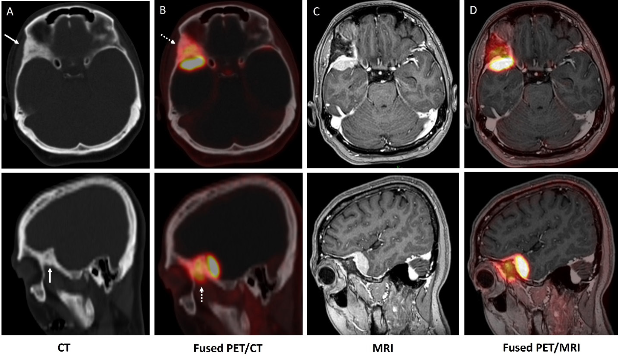
Meningiomas express the somatostatin receptor (SSTR). The utilization of SSTR ligands, specifically Gallium-68 (68Ga) isotope, a radioactive isotope (68Ga)-DOTA-labeled peptides, has demonstrated exceptional diagnostic precision for the detection of meningiomas, primarily due to the absence of normal brain and bone activity. We report a case of a 48-year-old woman with sphenoid wing meningioma who underwent 68Ga-DOTA TOC positron emission tomography (PET) for tumor delineation. 68Ga-DOTA TOC PET shows SSTR-avid meningioma in the right sphenoid/anterior temporal region with significant hyperostosis with high expression of SSTR in the bone. 68Ga-DOTA TOC uptake in the hyperostosis signifies bone infiltration rather than reactive changes. 68Ga-DOTA PET provides a better assessment of osseous involvement and provides additional information in terms of meningioma extent and planning for further management.
Keywords: 68Ga-DOTA, positron emission tomography/computed tomography, meningioma, SSTR, hyperostosis
,
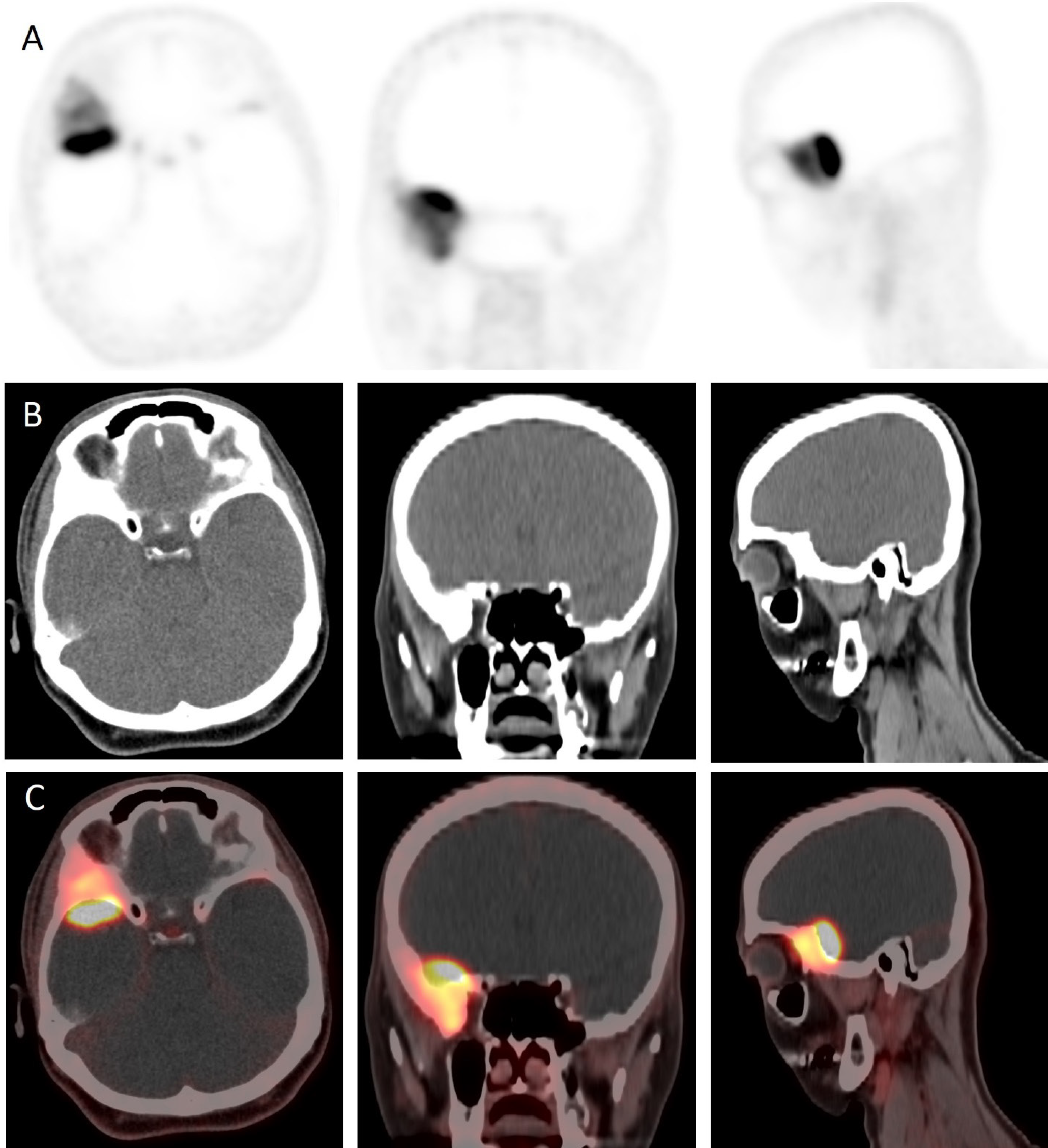
Meningiomas are the most common primary brain tumors, accounting for approximately 30% of intracranial tumors (). Bony involvement in intracranial meningiomas is uncommon and is believed to occur due to hyperostosis, which is reported to occur in 4.5%-17% of cases. Neoplastic infiltration of intracranial bone and primary intraosseous meningioma are other causative factors for bone involvement (). It is more frequently observed in meningiomas with plaque, with an occurrence rate of 13%-49% (). Transosseous extension of intracranial meningiomas is associated with a high risk of tumor recurrence and mortality and often requires gross total resection and cranial reconstruction (). There is no clear mechanism underlying the hyperostotic changes that have been described in the literature. Hyperostosis may occur due to irritation from an adjacent tumor, leading to bone invasion by meningiomatosis cells, resulting in secondary hypervascularity in the diploe of the surrounding skull. Some authors have suggested that the infiltration of bone by tumor cells causes secondary changes in osteoblast and osteoclast activity, leading to increased bone deposition. A small cohort study by Matschke J et al. () suggests that there is high expression of somatostatin receptor 2A (SSR2A) during the histogenesis of hyperostosis in meningioma. They found that the osseous part of the tumor exhibited the same strong SSR2A reactivity as the extraosseous part of the tumor, which possibly favors bone infiltration rather than reactive changes.
Somatostatin receptor (SSTR) is a 68G-protein-coupled receptor that is highly expressed in the majority of meningiomas, with subtype 2 being the most abundant (). 68Ga-DOTA PET imaging with radiolabeled somatostatin agonists offers high diagnostic accuracy for assessing meningiomas because of the high density of SSTRs. This modality provides additional information for patients exhibiting ambiguous or equivocal results on MRI. 68Ga-DOTA PET delineates tumor extent more accurately than MRI with contrast, particularly in regions such as the skull base, orbital area, and parasagittal regions, where bone invasion or involvement of the dural sinuses is frequently observed (). 68Ga-DOTA PET is also more proficient in detecting osseous involvement than MRI, as the detection of osseous infiltration relies on morphologic features such as hyperostosis and intraosseous contrast enhancement (). It is difficult to precisely define the degree of infiltration on CT and MRI images. 68Ga-DOTA PET provides a better assessment of osseous involvement and has great potential to improve target delineation for surgical resection and radiation treatment planning (). 68Ga-DOTA PET can accurately determine tumor margins for radiotherapy. A recent study by Kowalski ES et al. () reported the value of 68Ga-DOTATATE PET/CT in the diagnosis, radiation treatment planning, and evaluation of the response of meningiomas to radiotherapy.
Footnotes
Ethics
Informed Consent: Patient consent was not required for this study.
Authorship Contributions
Surgical and Medical Practices: S.F.H., J.K., Concept: S.U., A.J., K.A.R., S.F.H., S.K., Design: S.U., A.J., S.K., J.K., Data Collection or Processing: S.U., J.K., Analysis or Interpretation: S.U., S.F.H., A.J., K.A.R., S.K., Literature Search: S.U., Writing: S.U., A.J., K.A.R.
Conflict of Interest: No conflicts of interest were declared by the authors.
Financial Disclosure: The authors declare that this study has received no financial support
References
Ostrom QT, Gittleman H, Truitt G, Boscia A, Kruchko C, Barnholtz-Sloan JS. CBTRUS statistical report: Primary brain and other central nervous system tumors diagnosed in the united states in 2011-2015. Neuro Oncol. 2018;20(suppl_4):iv1-86.
Pieper DR, Al-Mefty O, Hanada Y, Buechner D. Hyperostosis associated with meningioma of the cranial base: secondary changes or tumor invasion. Neurosurgery. 1999;44:742-746
Akutsu H, Sugita K, Sonobe M, Matsumura A. Parasagittal meningioma en plaque with extracranial extension presenting diffuse massive hyperostosis of the skull. Surg Neurol. 2004;61:165-169.
Ildan F, Erman T, Göçer AI, Tuna M, Bağdatoğlu H, Cetinalp E, Burgut R. Predicting the probability of meningioma recurrence in the preoperative and early postoperative period: a multivariate analysis in the midterm follow-up. Skull Base. 2007;17:157-171.
Matschke J, Addo J, Bernreuther C, Zustin J. Osseous changes in meningioma en plaque. Anticancer Res. 2011;31:591-596.
Theodoropoulou M, Stalla GK. Somatostatin receptors: from signaling to clinical practice. Front Neuroendocrinol. 2013;34:228-252.
Rachinger W, Stoecklein VM, Terpolilli NA, Haug AR, Ertl L, Pöschl J, Schüller U, Schichor C, Thon N, Tonn JC. Increased 68Ga-DOTATATE uptake in PET imaging discriminates meningioma and tumor-free tissue. J Nucl Med. 2015;56:347-353.
Kunz WG, Jungblut LM, Kazmierczak PM, Vettermann FJ, Bollenbacher A, Tonn JC, Schichor C, Rominger A, Albert NL, Bartenstein P, Reiser MF, Cyran CC. Improved detection of transosseous meningiomas using 68Ga-DOTATATE PET/CT compared with contrast-enhanced MRI. J Nucl Med. 2017;58:1580-1587.
Perlow HK, Siedow M, Gokun Y, McElroy J, Matsui J, Zoller W, Beyer S, Arnett A, Blakaj D, Boulter D, Fritz J, Miller E, Raval R, Kleefisch C, Bovi J, Palmer JD. 68Ga-DOTATATE PET-Based Radiation Contouring Creates More Precise Radiation Volumes for Patients With Meningioma. Int J Radiat Oncol Biol Phys. 2022;113:859-865.
Kowalski ES, Khairnar R, Gryaznov AA, Kesari V, Koroulakis A, Raghavan P, Chen W, Woodworth G, Mishra M. 68Ga-DOTATATE PET-CT as a tool for radiation planning and evaluating treatment responses in the clinical management of meningiomas. Radiat Oncol. 2021;16:151.

