

Abstract
Objectives
Among the most important diagnostic indicators of colorectal cancer; however, measuring molecular alterations are invasive and expensive. This study aimed to investigate the application of image processing to predict molecular alterations in colorectal cancer.
Methods
In this scoping review, we searched for relevant literature by searching the Web of Science, Scopus, and PubMed databases. The method of selecting the articles and reporting the findings was according to the guidelines of the Preferred Reporting Items for Systematic Reviews and Meta-Analyses; moreover, the Strengthening the Reporting of Observational Studies in Epidemiology checklist was used to assess the quality of the studies.
Results
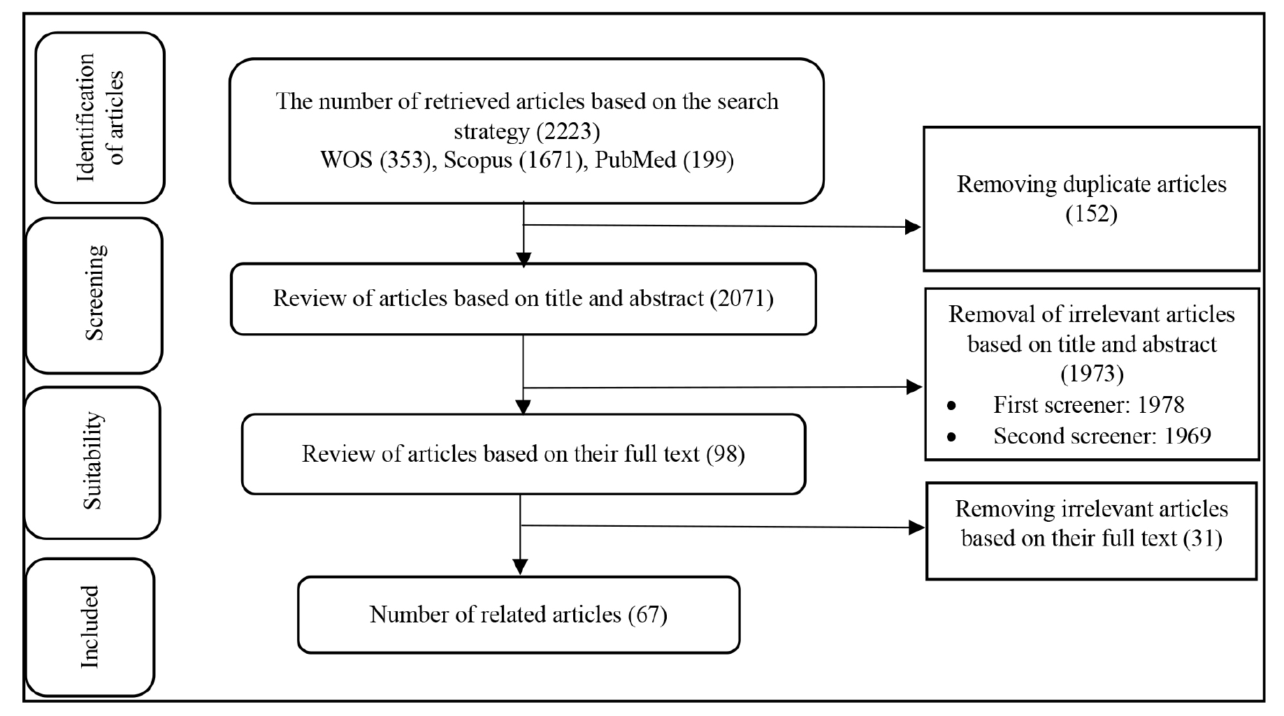
Sixty seven out of 2,223 articles, 67 were relevant to the aim of the study, and finally 41 studies with sufficient quality were reviewed. The prediction of Kirsten Rat Sarcoma Viral Oncogene Homolog (KRAS), Neuroblastoma RAS Viral (NRAS), B-Raf proto-oncogene, serine/threonine kinase (BRAF), Tumor Protein 53 (TP53), Adenomatous Polyposis Coli, and microsatellite instability (MSI) with the help of image analysis has received more attention than other molecular characteristics. The studies used computed tomography (CT), magnetic resonance imaging (MRI), and 18F-FDG positron emission tomography (PET)/CT with radionics and quantitative analysis to predict molecular alterations in colorectal cancer, analyzing features like texture, maximum standard uptake value, and MTV using various statistical methods. In 39 studies, there was a significant relationship between the features extracted from these images and molecular alterations. Different modalities were used to measure the area under the receiver operating characteristic curve for predicting the alterations in KRAS, MSI, BRAF, and TP53, with an average of 78, 81, 80 and 71%, respectively.
Conclusion
This scoping review underscores the potential of radiogenomics in predicting molecular alterations in colorectal cancer through non-invasive imaging modalities, like CT, MRI, and 18F-FDG PET/CT. The analysis of 41 studies showed the appropriate prediction of key alterations, such as KRAS, NRAS, BRAF, TP53, and MSI, highlighting the promise of radionics and texture features in enhancing predictive accuracy.
Keywords: Radiogenomics, colorectal cancer, molecular alterations, image processing
Introduction
Colorectal cancer is the third most common cancer in the world; it ranks second in men and third in women in terms of cancer-related deaths (, ). In 2022, a total of 1,926,118 new cancer cases and 903,859 deaths were reported (). Although the incidence of cancer has decreased in high-income countries because of continuous screenings in the elderly and changes in risk factors (, ), it is still increasing in low-income countries (, ).
Colorectal cancer, which is caused by the accumulation of genetic and epigenetic changes in the colon epithelium, is a complex heterogeneous disease with different histopathology, (). These changes lead to the activation of oncogenes, inactivation of tumor suppressor genes, and disturbance in the regulation of signaling pathways involved in cell proliferation, differentiation, and apoptosis (). As a result of different histopathology and heterogeneity, the progress of colorectal cancer is very different in different people. Therefore, it is very important to predict disease progression to determine the appropriate treatment (). To date, many efforts have been made to identify factors affecting disease progression, such as the “Tumor”, “Nodes”, “Metastases” (TNM) classification which, from histopathology point of view, classifies cancer into four groups with different rates of disease progression (, ). However, the rate of disease progression in the TNM groups differed due to the molecular differentiation and heterogeneity within the tumor ().
One-way to predict disease progression is to pay attention to molecular alterations, such as Kirsten Rat Sarcoma Viral Oncogene Homolog (KRAS), Neuroblastoma RAS Viral (NRAS), B-Raf proto-oncogene, serine/threonine kinase (BRAF), Tumor Protein 53 (TP53), microsatellite instability (MSI) and PIK3CA (). Current methods for measuring these factors in colorectal cancer, such as DNA sequence analysis, are costly, time-consuming, and invasive (). In addition, sampling from one point of the tumor to perform genetic tests and heterogeneity in different parts of the tumor, this method may not accurately reflect the molecular alterations of colorectal cancer (). The problems of measuring molecular alterations can be overcome by predicting their values through analyzing medical images, such as computed tomography (CT) scan, magnetic resonance imaging (MRI), and 18F-FDG PET/CT, which have recently attracted the attention of researchers (). Radiogenomics, a new concept introduced in recent years, examines the relationship between molecular alterations (especially genetic alterations) of cells and images (). Non-invasive imaging provides information on tumor morphology and metabolism to some extent and can be used to identify potential biomarkers and molecular alterations in colorectal cancer (). Several studies have shown that CT scanning can predict the KRAS mutation status in colorectal cancer patients (, ).
However, the use of image processing to predict MC molecular alterations is still in its early stages (, ). Therefore, this study aimed to investigate the use of image processing to predict molecular alterations in colorectal cancer. The research questions of this study are as follows:
1. Which molecular alterations have received more attention in the field of radiogenomics?
2. Which imaging modalities are used to predict molecular alterations?
3. What is the performances of the modalities in the prediction of various molecular alterations?
Materials and Methods
In this scoping review conducted in 2024, the reporting process was based on the Preferred Reporting Items for Systematic Reviews and Meta-Analysis (PRISMA) (). All original articles published from January 01, 2013 to April 31, 2024 and indexed in Scopus, PubMed, and Web of Science databases were extracted. The inclusion criteria were original research articles in colorectal cancer. The exclusion criteria were review studies, non-English articles, studies beyond the scope of colorectal cancer, and articles with limited access.
The standard keywords and their synonyms for the three terms “molecular alterations”, “medical image” and “colorectal cancer” were determined according to medical subject headings, and a search strategy was determined for each database (, ).
Table 1. Search strategy by database
Data base | Search strategy |
PubMed | ((((Gene[mesh] OR Genom*[Title/Abstract] OR “molecular alterations”[Title/Abstract] OR “Genes, APC”[mesh] OR “Genes, ras”[mesh] OR “Proto-Oncogene Proteins B-raf”[mesh] OR TP53 [Title/Abstract] OR “microsatellite instability”[mesh] OR MSI[Title/Abstract]) AND ( “Tomography, X-Ray Computed” [mesh] OR “Positron emission tomography” [mesh] OR “Magnetic resonance imaging” [mesh] OR “Diagnostic imaging” [mesh] OR Radiomics [mesh])) OR “radio-genomics” [Title/Abstract] OR radiogenomics [Title/Abstract] OR “imaging genomics” [Title/Abstract] OR “radiation genomics” [Title/Abstract]) AND (Colorectal Neoplasms [mesh] OR CRC[Title/Abstract])) AND 2013/01/01: 2024/04/28 [dp] |
Web of Science | ((((TS=(Gene) OR TS=(Genom*)OR TS =(“ molecular alterations”)OR TS=( Cistron*)OR TS=( Genetic)OR TS=( “Genes, APC”) OR TS=( “Genes, ras”) OR TS=( RAS) OR TS=( APC) OR TS=( APC) OR TS=( “Proto-Oncogene Proteins B-raf”) OR TS=( BRAF) OR TS=( BRAF) OR TS=( “tumor protein p53”) OR TS=( TP53) OR TS=( “microsatellite instability”) OR TS=( MSI) ) AND (TS=( “Diagnostic imaging”) OR TS=( “CT scan”) OR TS=( MRI) OR TS=( “18F-FDG PET/CT”) OR TS=( “computerized tomography”) OR TS=( “Positron emission tomography”) OR TS=( “Magnetic resonance imaging”) OR TS=( Radiomics))) OR (TS=( “radio-genomics”) OR TS=( radiogenomics) OR TS=( “imaging genomics”) OR TS=( “radiation genomics”)))AND (TS=( “Colorectal Neoplasms”) OR TS=( “Colorectal cancer”) OR TS=( CRC))) AND PY=(2013-2024) |
Scopus | (((TITLE-ABS-KEY(Gene) OR TITLE-ABS-KEY(Cistron*) OR TITLE-ABS-KEY(“molecular alterations”) OR TITLE-ABS-KEY(Genetic) OR TITLE-ABS-KEY(genom*) OR TITLE-ABS-KEY(“Genes, APC”) OR TITLE-ABS-KEY(APC) OR TITLE-ABS-KEY(“Genes, ras”) OR TITLE-ABS-KEY(RAS) OR TITLE-ABS-KEY(BRAF) OR TITLE-ABS-KEY(“Proto-Oncogene Proteins B-raf”) OR TITLE-ABS-KEY(“tumor protein p53”) OR TITLE-ABS-KEY(TP53) OR TITLE-ABS-KEY(“microsatellite instability”) OR TITLE-ABS-KEY(MSI)) AND (TITLE-ABS-KEY(“Diagnostic imaging”) OR TITLE-ABS-KEY(“CT scan”) OR TITLE-ABS-KEY(MRI) OR TITLE-ABS-KEY(“18F-FDG PET/CT”) OR TITLE-ABS-KEY(“computerized tomography”) OR TITLE-ABS-KEY(“Magnetic resonance imaging”) OR TITLE-ABS-KEY(“Positron emission tomography”) OR TITLE-ABS-KEY(Radiomics))) OR (TITLE-ABS-KEY(radiogenomics) OR TITLE-ABS-KEY(“radio-genomics”) OR TITLE-ABS-KEY(“imaging genomics”) OR TITLE-ABS-KEY(“radiation genomics”))) AND (TITLE-ABS-KEY(“Colorectal Neoplasms”) OR TITLE-ABS-KEY(“Colorectal cancer”) OR TITLE-ABS-KEY(CRC))) AND (PUBYEAR > 2013 AND PUBYEAR < 2024) |
Table 2. Characteristics of reviewed studies
Studies | Participants | Molecular factors | Image | Statistics | ||||
|---|---|---|---|---|---|---|---|---|
Author | Type | Country/Year | Participants | Aim | Gene Oncogene/Suppressor | Modality | Analyzing techniques (features) | Technique |
Lovinfosse et al. (19) | Retrospective study | Belgium/2016 | 151 | Predicting RAS mutation as an indicator of treatment | KRAS NRAS RAS | 18F-FDG PET/CT | Radiomics (texture features) | Spearman correlation coefficient |
Yang et al. (20) | Retrospective study | China\ 2021 | 42 | Investigating the association between EIF2S2 and 18F-FDG PET/CT | EIF2S2 GLUT1 | 18F-FDG PET/CT | Radiomics [SUVmax, SUVmean, total lesion glycolysis (TLG), and metabolic tumor volume (MTV)] | Spearman’s correlation coefficient |
Huang et al. (21) | Cohort study | Taiwan \ 2022 | 71 | Finding therapeutic targets using radiomic features | PECAM1 PRDM1 AIF1 IL10 ISG20 TLR8 | CT scan | Radiomics (1,037 radiomic feature) | Heatmap visualization and correlation coefficient |
Jo and Kim. (22) | Retrospective study | South Korea \ 2018 | 75 | Investigating the association between KRAS mutations and MRI-based radiologic findings | KRAS | MRI | Axial to longitudinal tumor length (ATL/LTL) | The Mann-Whitney U test |
Yang et al. (23) | Retrospective study | China \ 2017 | 61 | Prediction of KRAS/NRAS/BRAF mutations | KRAS NRAS BRAF | CT scan | Radiomics [shape features, gray-level histogram features, gray-level co-occurrence matrix (GLCM) features, gray-level run-length matrix (GLRLM) features] | t-test or the Mann-Whitney U test for continuous variables and the chi-square test for categorical variables |
Xu et al. (24) | Retrospective study | China \ 2019 | 158 | Prediction of KRAS mutation as a therapeutic indicator | KRAS | MRI | Radiomics (texture features) | Mann–Whitney U test The chi-square test or Fisher exact test. |
Li et al. (25) | Retrospective study | China \ 2021 | 368 | Prediction of the microsatellite instability (MSI) status | MLH1 MSH2 MSH6 PMS2 | CT scan | Radiomics (intensity histogram, gray level co-occurrence matrix, gray level run length matrix, neighbor intensity difference, and shape) | Logistic regression, Support vector machine (SVM), Random forest, Gradient boosting machine (GBM), Naive Bayes |
Crimì et al. (26) | Retrospective study | Italy \ 2022 | 47 | Prediction of the presence of specific genetic mutations associated with CRC | MSI | CT scan | Radiomics [derived from histogram, run length matrix (RLM), gray-level co-occurrence matrix (GLCM), gray-level run length matrix (GLRLM), and neighboring gray-level dependence matrix (NGLDM)] | Mann-Whitney U test and Bonferroni correction |
Cho et al. (27) | Retrospective study | Republic of Korea \ 2017 | 93 | Prediction of KRAS mutations | KRAS | 18F-FDG PET/CT | Quantitative Analysis (SUVmax, SUVmean, MTV, and TLG) | the χ2 test or Mann-Whitney U test |
Chen et al. (28) | Retrospective study | Taiwan \ 2013 | 121 | Prediction of KRAS mutations | KRAS | 18F-FDG PET/CT | SUVmax, SUVmean, MTV | using a Mann-Whitney U test and logistic regression analysis |
Yeo et al. (29) | Retrospective study | Republic of Korea \ 2015 | 46 | Whether DCE-MRI correlates with angiogenesis and the biological aggressiveness of rectal cancer | EGFR | MRI | Quantitative Analysis( Ktrans, kep, ve, and iAUC) | Student’s t-test and analysis of variance (ANOVA) |
Mao et al. (30) | Retrospective study | China\ 2019 | 49 | Prediction of KRAS mutations | KRAS | 18F-FDG PET/CT | Quantitative Analysis( SUVearly, SUVdelayed, DSUVmax) | Chi-square test or Mann–Whitney U test |
Taguchi et al. (31) | Retrospective study | Japan \ 2019 | 40 | Prediction of KRAS mutations | KRAS | CT scan | Quantitative Analysis [CT texture parameters (Skewness, Kurtosis, Entropy, Energy, Homogeneity,..) and 18F-FDG PET parameter) SUVmax)] | two-tailed independent t-test and the Mann-Whitney U test |
Zhong et al. (32) | Retrospective study | China \ 2022 | 1601 | Prognostic prediction model for colorectal cancer | KRAS | CT scan | Radiomics (radiomics features) | Unsupervised deconvolution analysis |
Wu et al. (33) | Cohort study | China \ 2019 | 279 | Predicting KRAS status in patients with colorectal cancer (CRC) | KRAS | CT scan | Radiomics and deep learningn (hand-crafted features and deep learning features) | using multivariable logistic regression analysis |
Seth et al. (34) | Retrospective study | Canada \ 2021 | 20 | Investigating the association between TTE and genetic mutations | APC ARIDA1 TP53 AKT1 ATM BRAF CTNNB1 EGFR FBXW7 KRAS NRAS PIK3 CAPTENSMAD | MRI | Quantitative Analysis [target tumor enhancement (weak and strong TTE)] | Kruskal-Wallis test and Mann-Whitney and/or t-test |
Cui et al. (35) | Retrospective study | China \ 2020 | 304 | prediction of KRAS mutation | KRAS | MRI | Radiomics (radiomics feature) | mainly consist of univariate statistical tests |
Cao et al. (36) | Retrospective study | China\ 2020 | 124 | prediction of KRAS mutation | KRAS | CT scan | Quantitative analysis( DESCT parameters Including the monochromatic CT value, iodine, water, and effective atomic number | logistic regression analysis |
Cui et al. (37) | Retrospective study | China \ 2019 | 148 | prediction of KRAS mutations | KRAS | MRI | Quantitative analysis (DKI and ADC) | Student’s t-test or Mann-Whitney U test and receiver operating characteristic (ROC) curve analysis |
Granata et al. (38) | Retrospective study | Italy \ 2020 | 106 | Identification of RAS mutation in colorectal liver metastasis | KRAS NRAS | MRI | Quantitative analysis (ADC-, IVIM- and DKI) | Wilcoxon-Mann-Whitney U tests for receiver operating characteristic (ROC) analyses |
Chen et al. (39) | Retrospective study | Taiwan \ 2015 | 103 | Investigating the association between genetic mutations and 18F-FDG PET in colorectal cancer (CRC). | TP53 KRAS APC BRAF PIK3CA | 18F-FDG PET/CT | Quantitative analysis (SUVmax, SUVmean, MTV, and TLG) | Mann-Whitney U test and logistic regression analysis |
Promsorn et al. (40) | Retrospective study | Thailand \ 2021 | 113 | prediction of KRAS mutation status | KRAS | CT scan | Qualitative and quantitative analyses (ATL, LTL, ATL/LTL, tumor location, gross tumor patterns, tumor margins, tumor enhancement patterns, T staging, regional lymph node metastasis, distant lymph node metastasis, or distal organ metastasis) | t test Mann-Whitney U test |
Liu et al. (41) | Retrospective study | China \ 2021 | 134 | Prediction of metastasis in colorectal cancer | ABCB1 TP53 ATM MYC | CT scan | Radiomics (radiomics feature) | Multivariable logistic regression analyses |
Popovic et al. (42) | Retrospective study | USA \ 2020 | 37 | Explore the predictive value of 2-[18F]FDG uptake according to KRAS mutation status | KRAS mutation | 2-[18F]FDG PET/C | quantitative analysis(SUV (HERMES), SUV (MIM), PVEC) | Student’s t test Wilcoxon rank-sum test Logistic regression and receiver operating characteristics (ROC) |
Krikelis et al. (43) | Retrospective study | Greece \ 2014 | 44 | prediction of KRAS mutation status | GLUT1 KRAS | 18F-FDG PET/CT | quantitative analyses ( SUVmax) | t-test Kruskal–Wallis test Fisher’s exact test of Spearman’s Rho |
Chen et al. (44) | Retrospective study | Taiwan \ 2019 | 74 | Investigating the association between genetic mutations and radionics in 18F-FDG PET/CT In colorectal cancer (CRC) | KRAS TP53 APC | 18F-FDG PET/CT | quantitative analyses ( SUVmax, MTV) | Receiver- operating characteristic (ROC) Mann-Whitney U test Spearman’s rank correlation coefficient |
Zhang et al. (45) | Retrospective study | China \ 2021 | 83 | Analyzing the association between MRI radiomic features and KRAS status in LARC patients. | KRAS NRAS BRAF | MRI | Radiomics (radiomics feature) | The least absolute shrinkage and selection operator (LASSO) regression |
Miles et al. (46) | prospective study | United Kingdom \ 2014 | 33 | Exploring the potential of multifunctional imaging in providing a KRAS signature | KRAS | PET/CT | quantitative analysis[18F-FDG uptake (18F-FDG maximum standardized uptake value (SUVmax)], CT texture (expressed as mean of positive pixels (MPP)] | CT Texture Analyses (CT TA) CT TA was performed using the following parameters: TexRAD Recursive decision tree Monte Carlo analysis |
He et al. (47) | Retrospective study | China \ 2020 | 157 | prediction of KRAS mutation status | KRAS | CT scan | Radiomics (radiomics feature) | LASSO regression- radiomics model using a random forest classifier (RFC) |
Shi et al. (48) | Retrospective study | China\ 2020 | 159 | predicting the RAS(KRAS and NRAS) and BRAF gene mutation statuses | KRAS NRAS BRAF | CT scan | Radiomics( Gray Level Co-occurrence Matrix (GLCM), gray level size zone matrix (GLSZM), Gray Level Run Length Matrix (GLRLM), neighboring Gray Tone Difference Matrix (NGTDM), and gray level dependence matrix (GLDM)) | Fisher’s exact test |
He et al. (49) | Retrospective study | China \ 2021 | 85 | association between KRAS/NRAS /BRAF mutations and metabolic parameters of pretreatment 18FFDG-PET/CT in colorectal cancer | KRAS NRAS BRAF | 18F-FDG PET/CT | quantitative analyses (SUVmax) | Mann–Whitney U test. |
Sh et al. (50) | prospective study | Egypt\ 2021 | 38 | correlation between 18F-FDG PET/CT imaging and KRAS expression in mCRC | KRAS mutational | 18F-FDG PET/CT | quantitative analysis (maximum standardized uptake value (SUVmax), total lesion glycolysis (TLG) and metabolic tumor volume (MTV)) | independent samples t-test Mann-Whitney U test) |
Li et al. (51) | Retrospective study | China\ 2021 | 173 | Prediction of Microsatellite instability in colorectal tissue Cancer | MSI-H MSS | 18F-FDG PET/CT | Radiomics (radiomics feature) | multivariate random forest selection and univariate relevancy tests Balanced Bagging the area under The curve (AUC) |
Horvat et al. (52) | Retrospective study | USA\ 2019 | 65 | To investigate associations between genetic mutations and qualitative and quantitative features on magnetic resonance imaging (MRI) in rectal adenocarcinoma | APC TP53 KRAS PIK3CA BRCA2 ATM SOX9 FLT4 | MRI | Quantitative and qualitative analyses [Tumor Localization, Tumor Length (cm), Mucin Content, CRM distance (mm), CRM status, DWI restriction, Early perfusion on DCE, Metastatic lymph nodes] | Fisher’s exact test and Wilcoxon rank sum test |
Badic et al. (53) | Retrospective study | Franche\ 2019 | 64 | the relationship between imaging radiomic features and gene expression changes | ABCB1 ABCC2 ABCG2 ALDH1A1 CD166 (ALCAM) CDKN1A INHBB | CT scan | Radiomics [Flatness, Sum entropy (SENTR), entropy from Gray-level-co-occurrence-matrix (EntropyGLCM E), Gray-level non-uniformity (GLNUL)] | Kruskal-Wallis test |
Ma et al. (54) | Retrospective study | USA\ 2022 | 230 | explore whether the preoperative CT radionics can predict Status of microsatellite instability (MSI) in colorectal cancer (CRC) | LH1, MSH2 MSH6 PMS2 | CT scan | Radiomics ( first-order statistics, shape, gray-level co-occurrence matrix (GLCM), gray-level dependence matrix (GLDM), gray-level size zone matrix (GLSZM), gray-level run length matrix (GLRLM), and neighboring gray-level tone difference matrix (NGTDM)) | inter-class correlation coefficient intraclass correlation coefficient |
Negreros-Osuna et al. (55) | Retrospective study | Mexico\ 2020 | 145 | To explore the potential of radionics texture features as biomarkers of BRAF mutation | BRAF | CT scan | Radiomics (texture features: mean, SD, mean value of positive pixels (MPP), skewness, kurtosis, and entropy) | Laplacian-of-Gaussian filters Wilcoxon rank sum |
Granata et al. (56) | Retrospective study | Italy\ 2021 | 52 | Investigating the association between RAS mutation status and radiomics-derived data using contrast-enhanced magnetic resonance imaging (CE-MRI) in liver metastases | KRAS NRAS | MRI | Radiomics (texture features) | Wilcoxon-Mann-Whitney U Test Receiver Operating Characteristic (ROC) |
Kawada et al. (57) | Retrospective study | Japan\ 2015 | 55 | to investigate whether KRAS status is associated with 18F-FDG accumulation in metastatic CRC. and whether 18F-FDG PET/CT can be used to predict KRAS status of metastatic CRC. | KRAS | 18F-FDG PET/CT | quantitative analysis(SUVmax) | Mann–Whit- ney U test |
Mao et al. (30) | Retrospective study | China\ 2018 | 49 | To investigate the association between meta- Bolic parameters of dual time point 18 F-FDG PET/CT and KRAS mutation status in colorectal liver metastases (CRLM). | KRAS | 18F-FDG PET/CT | quantitative analysis (SUVearly, SUVdelayed, DSUVmax and RI) | Uni-variate multi-variate analyses |
Arslan et al. (58) | Retrospective study | Turkey\ 2020 | 83 | Investigating the association between FDG uptake patterns and 18 F-FDG PET/CT imaging and KRAS mutation | KRAS | 18F-FDG PET/CT | quantitative analyses (SUVmax) | Mann-Whitney U Kruskal-Wallis tests Pearson’s test. |
KRAS: Kirsten Rat Sarcoma Viral Oncogene Homolog, NRAS: Neuroblastoma RAS Viral, EIF2S2: Eukaryotic Translation Initiation Factor 2 Subunit 2 GLUTI: Glucose Box 1, BRAF: B-Raf proto-onkogen, MSI: Microsatellite Instability, PECAMI: Platelet Endothelial Cell Adhesion Molecule 1, AIFI: Activated Inducible Family of Immune receptors, ISG20: Interferon-Stimulated Gene 20, TLR8: Toll-like Receptor 8, CDKN2A: Cyclin-Dependent Kinase Inhibitor 2A, ATM: Ataxia-Telangiectasia Mutated, ABCG2: ATP Binding Cassette Subfamily G Member 2, ABCB1: ATP Binding Cassette Subfamily B Member 1, AMCC2: Armadillo Motif Containing 2, ABCG2: ATP Binding Cassette Subfamily G Member 2, ALDHIA1: Aldehyde Dehydrogenase 1 Family Member A1, CD166: Cluster of Differentiation 166, INHBB: Inhibin Beta B, CDKNIA: Cyclin-Dependent Kinase Inhibitor 1A, ARIDA1: AT-rich interaction domain 1A, CTNNBI: Catenin Beta 1, FBXW7: F-box and WD-40 domain protein 7, BRAC2: Breast Cancer 2, FLT: Fms-like Tyrosine Kinase
After searching and retrieving sources based on the search strategies, duplicate articles were removed using EndNote software. Then, the titles and abstracts of the articles were checked, and irrelevant articles were removed. This screening was performed by two experts in the field of Medical Informatics, and any disagreement was resolved by consensus with a third expert. In the next step, the full texts of the articles were reviewed. Finally, articles that were in line with the purpose of the study were selected. The quality of the selected articles was measured using the Strengthening the Reporting of Observational Studies in Epidemiology checklist, and articles with insufficient quality were excluded from the study ().
The data collection tool consisted of a data extraction form including the type of study, first author’s name, country and year of publication, purpose of the study, sample size, molecular factors, type of modality, image characteristics, statistical method for prediction, and summary of findings. Furthermore, a narrative synthesis method was used for data analysis.
Statistical Analysis
In this study, basic descriptive statistics, including the sum and mean, were used to analyze the results. Additionally, when the area under the ROC curve was calculated to predict molecular alterations in the studies, the weighted average was computed based on the molecular alterations and modality.
Patient Consent Information
This systematic review was based on data from previously published studies, and new patient data were not collected. Therefore, patient consent was not required.
Results
A summary of the study review process based on the PRISMA guidelines is presented in .
Molecular Factors
Research has shown that many molecular factors contribute to the treatment of colorectal cancer. Some of the key molecular factors (genes/oncogene/suppressor) in colorectal cancer are KRAS, BRAF, NRAS, PIK3CA, and TP53 (). Various molecular factors were predicted in the selected studies; however, in 68% of them, KRAS changes were investigated. The frequency of the investigated molecular factors is shown in .
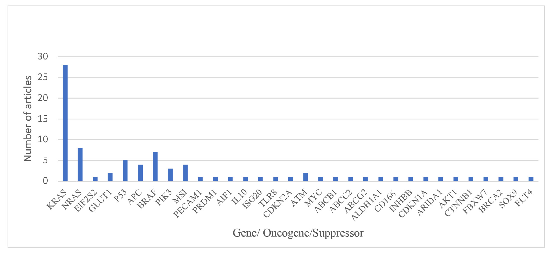
KRAS: Kirsten Rat Sarcoma Viral Oncogene Homolog, NRAS: Neuroblastoma RAS Viral, EIF2S2: Eukaryotic Translation Initiation Factor 2 Subunit 2 GLUTI: Glucose Box 1, BRAF: B-Raf proto-onkogen, MSI: Microsatellite Instability, PECAMI: Platelet Endothelial Cell Adhesion Molecule 1, AIFI: Activated Inducible Family of Immune receptors, ISG20: Interferon-Stimulated Gene 20, TLR8: Toll-like Receptor 8, CDKN2A: Cyclin-Dependent Kinase Inhibitor 2A, ATM: Ataxia-Telangiectasia Mutated, ABCG2: ATP Binding Cassette Subfamily G Member 2, ABCB1: ATP Binding Cassette Subfamily B Member 1, AMCC2: Armadillo Motif Containing 2, ABCG2: ATP Binding Cassette Subfamily G Member 2, ALDHIA1: Aldehyde Dehydrogenase 1 Family Member A1, CD166: Cluster of Differentiation 166, INHBB: Inhibin Beta B, CDKNIA: Cyclin-Dependent Kinase Inhibitor 1A, ARIDA1: AT-rich interaction domain 1A, CTNNBI: Catenin Beta 1, FBXW7: F-box and WD-40 domain protein 7, BRAC2: Breast Cancer 2, FLT: Fms-like Tyrosine Kinase
Modalities
Recently, different imaging modalities have been used for predicting molecular factors in colorectal cancer. The most important modalities are MRI, CT, and positron emission tomography (PET) (). The frequency of modalities used in the included studies is presented in .
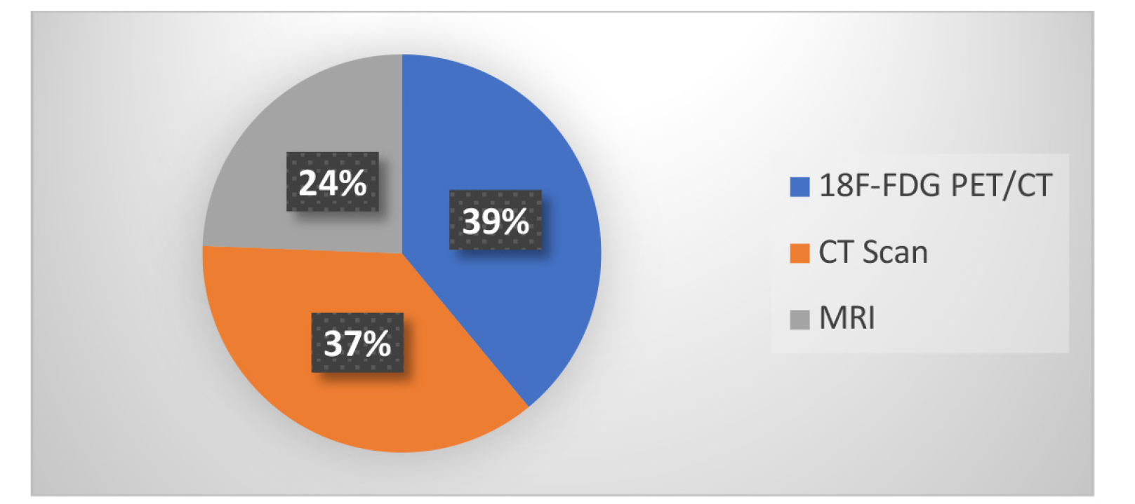
PET/CT: Positron emission tomography/computed tomography, MRI: Magnetic resonance imaging
Analyzing Technics
The reviewed studies employed various imaging modalities, such as CT, MRI, and 18F-FDG PET/CT, and utilized radionics and quantitative analysis techniques to predict molecular alterations in colorectal cancer. Key features analyzed included texture features, maximum standardized uptake value (SUVmax), SUVmean, metabolic tumor volume, total lesion glycolysis, and various radionics features derived from intensity, shape, and texture matrices like GLCM, GLRLM, GLSZM, and NGLDM. The statistical methods varied, including Spearman correlation, Mann-Whitney U test, logistic regression, and machine learning models like random forest and SVM.
Area Under the Receiver Operating Characteristic (ROC) Curve for Predicting Molecular Factors Based on Image
In 20 studies, the area under the ROC curve (AUC) was reported for predicting KRAS (n=13), MSI (n=4), BRAF (n=2), and TP53 (n=1) changes. The weighted average of this index (relative to the number of samples) for each molecular factor is presented in . presents the relationships among the three modalities used in studies on molecular factors.
Table 3. Area under the receiver operating characteristic curve of different modalities for predicting molecular factors
Molecular factors | Modality | Number of articles | Average sample size (SD of sample size) | AUC mean* |
|---|---|---|---|---|
KRAS | MRI | 5 | 153.6 (82.2) | 0.77 |
CT scan | 4 | 120 (48.23) | 0.82 | |
PET/CT | 4 | 82.5 (48.9) | 0.73 | |
ALL | 13 | 121.4 (68.5) | 0.78 | |
MSI | MRI | 0 | 0 (0) | 0 |
CT scan | 3 | 215 (131) | 0.80 | |
PET/CT | 1 | 173 (0) | 0.83 | |
ALL | 4 | 194 (115.4) | 0.81 | |
BRAF | MRI | 1 | 159 (0) | 0.79 |
CT scan | 1 | 61 (0) | 0.83 | |
PET/CT | 0 | 0 (0) | 0 | |
ALL | 2 | 110 (0) | 0.80 | |
TP53 | MRI | 0 | 0 (0) | 0 |
CT scan | 0 | 0 (0) | 0 | |
PET/CT | 1 | 74 (0) | 0.71 | |
ALL | 1 | 74 (0) | 0.71 |
*AUC mean, SD: Standard deviation, AUC: Area under the curve, MRI: Magnetic resonance imaging, CT: Computed tomography, PET: Positron emission tomography, ALL: Acute lymphoblastic leukemia, MSI: Mikrosatellit Instabilitesi, BRAF: B-Raf proto-oncogene, serine/threonine kinase, TP53: Tumor Protein 53
Table 4. Relationship between molecular alterations and image quality by modality
Modality | Gene | Positive* | Negative** | Total | ||
|---|---|---|---|---|---|---|
Number of articles | Average sample size (SD of sample size) | Number of articles | Average of sample size (SD of sample size) | Number of articles | ||
18F-FDG PET/CT | KRAS | 12 | 78.17 (34.24) | 1 | 44 (0) | 13 |
NRAS | 2 | 118 (33) | 0 | 0 (0) | 1 | |
EIF2S2 | 1 | 42 (0) | 0 | 0 (0) | 2 | |
GLUT1 | 1 | 42 (0) | 1 | 44 (0) | 2 | |
TP53 | 2 | 88.5 (14.5) | 0 | 0 (0) | 2 | |
APC | 1 | 74 (0) | 1 | 103 (0) | 2 | |
BRAF | 1 | 85 (0) | 1 | 103 (0) | 1 | |
PIK3CA | 0 | 0 (0) | 1 | 103 (0) | 1 | |
MSI | 1 | 173 (0) | 0 | 0 (0) | 1 | |
CT scan | PECAM1 | 1 | 71 (0) | 0 | 0 (0) | 1 |
PRDM1 | 1 | 71 (0) | 0 | 0 (0) | 1 | |
AIF1 | 1 | 71 (0) | 0 | 0 (0) | 1 | |
IL10 | 1 | 71 (0) | 0 | 0 (0) | 1 | |
ISG20 | 1 | 71 (0) | 0 | 0 (0) | 1 | |
TLR8 | 1 | 71 (0) | 0 | 0 (0) | 6 | |
KRAS | 6 | 109 (43.1) | 0 | 0 (0) | 2 | |
NRAS | 2 | 110 (49) | 0 | 0 (0) | 3 | |
BRAF | 3 | 121.6 (43.33) | 0 | 0 (0) | 3 | |
MSI | 3 | 215 (131) | 0 | 0 (0) | 1 | |
CDKN2A | 1 | 134 (0) | 0 | 0 (0) | 1 | |
TP53 | 1 | 134 (0) | 0 | 0 (0) | 1 | |
ATM | 1 | 134 (0) | 0 | 0 (0) | 1 | |
MYC | 1 | 134 (0) | 0 | 0 (0) | 1 | |
ABCB1 | 1 | 64 (0) | 0 | 0 (0) | 1 | |
ABCC2 | 1 | 64 (0) | 0 | 0 (0) | 1 | |
ABCG2 | 1 | 64 (0) | 0 | 0 (0) | 1 | |
ALDH1A1 | 1 | 64 (0) | 0 | 0 (0) | 1 | |
CD166 | 1 | 64 (0) | 0 | 0 (0) | 1 | |
INHBB | 1 | 64 (0) | 0 | 0 (0) | 1 | |
CDKN1A | 1 | 64 (0) | 0 | 0 (0) | 1 | |
MRI | KRAS | 8 | 118.2 (86.8) | 1 | 65 (0) | 9 |
EGFR | 2 | 33 (13.2) | 0 | 0 (0) | 2 | |
APC | 1 | 20 (0) | 1 | 65 (0) | 2 | |
ARIDA1 | 1 | 20 (0) | 0 | 0 (0) | 1 | |
TP53 | 2 | 42.5 (22.5) | 0 | 0 (0) | 2 | |
AKT1 | 1 | 20 (0) | 0 | 0 (0) | 1 | |
ATM | 2 | 42.5 (22.5) | 0 | 0 (0) | 2 | |
BRAF | 1 | 20 (0) | 1 | 83 (0) | 2 | |
CTNNB1 | 1 | 20 (0) | 0 | 0 (0) | 1 | |
FBXW7 | 1 | 20 (0) | 0 | 0 (0) | 1 | |
NRAS | 3 | 65.4 (36.3) | 1 | 65 (0) | 4 | |
PIK3 | 1 | 20 (0) | 1 | 65 (0) | 2 | |
BRCA2 | 0 | 0 (0) | 1 | 65 (0) | 1 | |
SOX9 | 0 | 0 (0) | 1 | 65 (0) | 1 | |
FLT4 | 0 | 0 (0) | 1 | 65 (0) | 1 | |
*Positive=There is a correlation between image features and molecular alterations, **Negetive=There is no correlation/relationship between image features and molecular alterations, KRAS: Kirsten Rat Sarcoma Viral Oncogene Homolog, NRAS: Neuroblastoma RAS Viral, EIF2S2: Eukaryotic Translation Initiation Factor 2 Subunit 2 GLUTI: Glucose Box 1, BRAF: B-Raf proto-onkogen, MSI: Microsatellite Instability, PECAMI: Platelet Endothelial Cell Adhesion Molecule 1, AIFI: Activated Inducible Family of Immune receptors, ISG20: Interferon-Stimulated Gene 20, TLR8: Toll-like Receptor 8, CDKN2A: Cyclin-Dependent Kinase Inhibitor 2A, ATM: Ataxia-Telangiectasia Mutated, ABCG2: ATP Binding Cassette Subfamily G Member 2, ABCB1: ATP Binding Cassette Subfamily B Member 1, AMCC2: Armadillo Motif Containing 2, ABCG2: ATP Binding Cassette Subfamily G Member 2, ALDHIA1: Aldehyde Dehydrogenase 1 Family Member A1, CD166: Cluster of Differentiation 166, INHBB: Inhibin Beta B, CDKNIA: Cyclin-Dependent Kinase Inhibitor 1A, ARIDA1: AT-rich interaction domain 1A, CTNNBI: Catenin Beta 1, FBXW7: F-box and WD-40 domain protein 7, BRAC2: Breast Cancer 2, FLT: Fms-like Tyrosine Kinase
Discussion
In this systematic review, 41 studies related to the use of radiogenomics in colorectal cancer for predicting molecular factors were examined. According to the results, 42% of the studies were conducted in China, and 71% of the studies were conducted between 2019 and 2022. According to recent progress in understanding the relationship between molecular factors and response to drugs, the emergence of the concept of radiogenomics, and the increase in the quality of different modalities in recent years, such studies have received much attention from researchers ().
Furthermore, most studies have investigated the association between medical images and RAS (KRAS, NRAS), BRAF, TP53, activated protein C (APC), and MSI alterations with a frequency of 35, 28, 8, 7, 5, 4, and 4, in that order. RAS mutations (KRAS/NRAS) are common in colorectal cancer and can affect treatment outcomes. These mutations are associated with resistance to anti-epidermal growth factor receptor monoclonal antibodies and limit their efficacy. Targeted therapies that specifically inhibit mutant KRAS are being developed to overcome this resistance (, ). RAS mutations have been investigated in 35 studies, and the relationship between image characteristics and molecular factors was significant in 33 studies. Moreover, 13 studies used image analysis to report the area under the ROC curve for predicting KRAS whose weighted average, relative to the number of samples, was 78%, which is in contrast with the result of the study by Kim et al. (), where the same value for 9 studies was 69%. This difference can be attributed to the research period. Additionally, regarding recent advances in imaging and image-analyzing methods, the higher level under the ROC curve in the present study can be justified.
BRAF mutations, particularly V600E mutation, are found in a subset of colon cancers and are associated with poor prognosis. In recent years, BRAF inhibitors have shown promise in the treatment of colorectal cancer (with BRAF mutation), either alone or in combination with other drugs (). BRAF mutation has been examined in 7 studies, and the relationship between image features and BRAF mutation was significant in 6 studies. In 2 studies image analyzing was used to report the area under the ROC curve for predicting BRAF whose weighted average, in relation to the number of samples, was 80%. In their study, Santhanam et al. () identified 7 studies on the relationship between 18F-FDG PET/CT characteristics and BRAF mutation in thyroid cancer, and the results indicated a significant relationship between them.
TP53 is a tumor suppressor that plays an important role in maintaining genomic stability. TP53 mutations are frequently found in colorectal cancer, and they are associated with worse prognosis and resistance to therapy. New therapies targeting TP53 mutations (such as gene therapies and small molecule inhibitors) are being investigated to overcome these challenges (). The TP53 mutation has been investigated in 5 studies where the relationship between image characteristics and TP53 mutation was significant. In another study, image analysis was used, and the area under the receiver operating characteristic curve for predicting TP53 was 71%. In their review study, Seow et al. () investigated the relationship between radiomic features and molecular factors and found a correlation between TP53 mutation and radiomic features.
MSI is observed in approximately 15% of colorectal cancers. From the treatment point of view, high MSI colorectal cancers exhibit particular responses to immunotherapy; they respond better to immune checkpoint inhibitors (). Image analysis was used in 4 studies to report the area under the receiver operating characteristic curve for MSI, with an average of 81%. Similarly, Le et al. () identified 8 studies related to the use of radionics for the prediction of MSI, and the average area under the ROC curve was 83%.
As a suppressor, APC plays an important role in the development of colon cancer and is used to identify people at risk or diagnose the disease. In addition, Wnt pathway inhibitors therapies may be appropriate for APC-mutated colorectal cancer (). APC mutation has been examined in 4 studies in which the relationship between image feature and APC changes was significant. A review study by Aghabozorgi et al. (), on the relationship between radionics features and histopathological changes indicated a relationship between APC mutation and radionics features.
MRI, CT scanning, and 18F-FDG PET/CT were used in 10, 15, and 16 studies, respectively. MRI is a non-invasive imaging technique that provides high-resolution anatomical images. It provides good soft-tissue contrast and is useful for evaluating colorectal tumor characteristics, such as size, location, and invasion depth (, ). According to the performance of MRI, this modality is mostly used for predicting RAS (KRAS/NRAS).
There was also a significant relationship between MRI and molecular factors in all selected studies, except for Horvat et al. () study in which qualitative characteristics of images were related to molecular factors; however, no significant relationship was found between quantitative characteristics and molecular factors due to the limitations presented in the study.
The analysis of radionics and quantitative features across various imaging modalities, such as CT, MRI, and 18F-FDG PET/CT, revealed the potential for predicting molecular alterations in colorectal cancer. Radiomics features, including texture and intensity metrics, can help improve the prediction accuracy. Techniques like GLCM, GLRLM, and GLSZM combined with statistical methods such as logistic regression and machine learning models demonstrate varying degrees of success in identifying key genetic mutations such as KRAS, NRAS, BRAF, TP53, and MSI. However, the heterogeneity in methodologies and sample sizes across studies underscores the need for standardized imaging protocols and radiomic analysis techniques.
Study Limitations
Because the studies were conducted considering a small sample size and were still in their early stages, multi-center prospective studies with a larger number of participants should be conducted.
Conclusion
This scoping review highlights the promising potential of radiogenomics in predicting molecular alterations in colorectal cancer through noninvasive imaging modalities. Our comprehensive analysis of 41 high-quality studies revealed that various imaging techniques, including CT scanning, MRI, and 18F-FDG PET/CT, can effectively predict key molecular changes, such as KRAS, NRAS, BRAF, TP53, and MSI. The primary focus has been on CT scanning and MRI, with texture features and radionics playing critical roles in enhancing predictive accuracy. Despite these advancements, the field is still in its nascent stages, with varying levels of predictive performance and sample sizes. The heterogeneity of methodologies and the need for larger, more diverse cohorts underscore the need for further research. Standardization of imaging protocols and radiomic analysis, along with cross-institutional collaborations, will be crucial for validating and refining these predictive models. In conclusion, radiogenomics has significant potential to revolutionize the prediction of molecular alterations in colorectal cancer, facilitating personalized treatment approaches. Continued research and technological advancements are essential for fully realizing its clinical implications and improving patient outcomes.
Footnotes
Ethics
Ethics Committee Approval: This study did not require ethical approval as it did not involve interaction with patients or human subjects.
Informed Consent: This systematic review was based on data from previously published studies, and new patient data were not collected. Therefore, patient consent was not required.
Authorship Contributions
Concept: S.M., H. E., R.R., Design: S.M., H. E., R.R., Data Collection or Processing: S.M., H.M., F.F., Analysis or Interpretation: S.M., H. E., A.H., Literature Search: S.M., A.H., H.M., F.F., R.B., Writing: S.M., H. E., R.R., R.B.
Conflict of Interest: No conflict of interest was declared by the authors.
Financial Disclosure: The authors declared that this study has received no financial support.
References
Patel SG, Karlitz JJ, Yen T, Lieu CH, Boland CR. The rising tide of early-onset colorectal cancer: a comprehensive review of epidemiology, clinical features, biology, risk factors, prevention, and early detection. Lancet Gastroenterol Hepatol. 2022;7:262-274.
Kim SJ, Pak K, Kim K. Diagnostic performance of F-18 FDG PET/CT for prediction of KRAS mutation in colorectal cancer patients: a systematic review and meta-analysis. Abdom Radiol. 2019;44:1703-1711.
Bray F, Laversanne M, Sung H, Ferlay J, Siegel RL, Soerjomataram I, Jemal A. Global cancer statistics 2022: GLOBOCAN estimates of incidence and mortality worldwide for 36 cancers in 185 countries. CA Cancer J Clin. 2024;74:229-263.
Rabiei R, Ayyoubzadeh SM, Sohrabei S, Esmaeili M, Atashi A. Prediction of breast cancer using machine learning approaches. J Biomed Phys Eng. 2022;12:297-308.
Awedew AF, Asefa Z, Belay WB. Burden and trend of colorectal cancer in 54 countries of Africa 2010-2019: a systematic examination for Global Burden of Disease. BMC Gastroenterol. 2022;22:204.
Mehdizadeh H, Asadi F, Mehrvar A, Nazemi E, Emami H. Smartphone apps to help children and adolescents with cancer and their families: a scoping review. Acta Oncol. 2019;58:1003-1014.
Sveen A, Kopetz S, Lothe RA. Biomarker-guided therapy for colorectal cancer: strength in complexity. Nat Rev Clin Oncol. 2020;17(1):11-32.
Vogelstein B, Papadopoulos N, Velculescu VE, Zhou S, Diaz Jr LA, Kinzler KW. Cancer genome landscapes. Science. 2013;339:1546-1558.
Badic B, Tixier F, Cheze Le Rest C, Hatt M, Visvikis D. Radiogenomics in colorectal cancer. Cancers. 2021;13:973.
Siegel RL, Miller KD, Goding Sauer A, Fedewa SA, Butterly LF, Anderson JC, Cercek A, Smith RA, Jemal A. Colorectal cancer statistics, 2020. CA Cancer J Clin. 2020;70:145-164.
Dagogo-Jack I, Shaw AT. Tumour heterogeneity and resistance to cancer therapies. Nat Rev Clin Oncol. 2018;15:81-94.
Liu Z, Zhang XY, Shi YJ, Wang L, Zhu HT, Tang Z, Wang S, Li XT, Tian J, Sun YS. Radiomics analysis for evaluation of pathological complete response to neoadjuvant chemoradiotherapy in locally advanced rectal cancer. Clin Cancer Res. 2017;23:7253-7262.
Burrell RA, Swanton C. Tumour heterogeneity and the evolution of polyclonal drug resistance. Mol Oncol. 2014;8:1095-1111.
Li M, Zhang J, Dan Y, Yao Y, Dai W, Cai G, Yang G, Tong T. A clinical-radiomics nomogram for the preoperative prediction of lymph node metastasis in colorectal cancer. J Transl Med. 2020;18:46.
Liu S, Pan X, Liu R, Zheng H, Chen L, Guan W, Wang H, Sun Y, Tang L, Guan Y, Ge Y, He J, Zhou Z. Texture analysis of CT images in predicting malignancy risk of gastrointestinal stromal tumours. Clin Radiol. 2018;73:266-274.
de la Pinta C, Castillo ME, Collado M, Galindo-Pumariño C, Peña C. Radiogenomics: hunting down liver metastasis in colorectal cancer patients. Cancers. 2021;13:5547.
Page MJ, McKenzie JE, Bossuyt PM, Boutron I, Hoffmann TC, Mulrow CD, Shamseer L, Tetzlaff JM, Akl EA, Brennan SE, Chou R, Glanville J, Grimshaw JM, Hróbjartsson A, Lalu MM, Li T, Loder EW, Mayo-Wilson E, McDonald S, McGuinness LA, Stewart LA, Thomas J, Tricco AC, Welch VA, Whiting P, Moher D. The PRISMA 2020 statement: an updated guideline for reporting systematic reviews. Int J Surg. 2021;88:105906.
Darvish L, Bahreyni-Toossi M-T, Roozbeh N, Azimian H. The role of radiogenomics in the diagnosis of breast cancer: a systematic review. Egypt J Med Hum Genet. 2022;23:1-16.
Lovinfosse P, Koopmansch B, Lambert F, Jodogne S, Kustermans G, Hatt M, Visvikis D, Seidel L, Polus M, Albert A, Delvenne P, Hustinx R. 18F-FDG PET/CT imaging in rectal cancer: relationship with the RAS mutational status. Br J Radiol. 2016;89:20160212.
Yang JW, Yuan LL, Gao Y, Liu XS, Wang YJ, Zhou LM, Kui XY, Li XH, Ke CB, Pei ZJ. 18F-FDG PET/CT metabolic parameters correlate with EIF2S2 expression status in colorectal cancer. J Cancer. 2021;12:5838-5847.
Huang YC, Tsai YS, Li CI, Chan RH, Yeh YM, Chen PC, Shen MR, Lin PC. Adjusted CT image-based radiomic features combined with immune genomic expression achieve accurate prognostic classification and identification of therapeutic targets in stage III colorectal cancer. 2022;14:1895.
Jo SJ, Kim SH. Association between oncogenic RAS mutation and radiologic-pathologic findings in patients with primary rectal cancer. Quant Imaging Med Surg. 2019;9:238-246.
Yang L, Dong D, Fang M, Zhu Y, Zang Y, Liu Z, Zhang H, Ying J, Zhao X, Tian J. Can CT-based radiomics signature predict KRAS/NRAS/BRAF mutations in colorectal cancer? Eur Radiol. 2018;28:2058-2067.
Xu Y, Xu Q, Ma Y, Duan J, Zhang H, Liu T, Li L, Sun H, Shi K, Xie S, Wang W. Characterizing MRI features of rectal cancers with different KRAS status. BMC cancer. 2019;19:1111.
Li Z, Zhong Q, Zhang L, Wang M, Xiao W, Cui F, Yu F, Huang C, Feng Z. Computed tomography-based radiomics model to preoperatively predict microsatellite instability status in colorectal cancer: a multicenter study. Front Oncol. 2021;11:666786.
Crimì F, Zanon C, Cabrelle G, Luong KD, Albertoni L, Bao QR, Borsetto M, Baratella E, Capelli G, Spolverato G, Fassan M, Pucciarelli S, Quaia E. Contrast-enhanced CT texture analysis in colon cancer: correlation with genetic markers. Tomography. 2022;8:2193-2201.
Cho A, Jo K, Hwang SH, Lee N, Jung M, Yun M, Hwang HS. Correlation between KRAS mutation and 18 F-FDG uptake in stage IV colorectal cancer. Abdom Radiol. 2017;42:1621-1626.
Chen SW, Chiang HC, Chen WT, Hsieh TC, Yen KY, Chiang SF, Kao CH. Correlation between PET/CT parameters and KRAS expression in colorectal cancer. Clin Nucl Med. 2014;39:685-689.
Yeo DM, Oh SN, Jung CK, Lee MA, Oh ST, Rha SE, Jung SE, Byun JY, Gall P, Son Y. Correlation of dynamic contrast‐enhanced MRI perfusion parameters with angiogenesis and biologic aggressiveness of rectal cancer: preliminary results. J Magn Reson Imaging. 2015;41:474-480.
Mao W, Zhou J, Zhang H, Qiu L, Tan H, Hu Y, Shi H. Relationship between KRAS mutations and dual time point 18 F-FDG PET/CT imaging in colorectal liver metastases. Abdominal Radiology. 2019;44:2059-2066.
Taguchi N, Oda S, Yokota Y, Yamamura S, Imuta M, Tsuchigame T, Nagayama Y, Kidoh M, Nakaura T, Shiraishi S, Funama Y, Shinriki S, Miyamoto Y, Baba H, Yamashita Y. CT texture analysis for the prediction of KRAS mutation status in colorectal cancer via a machine learning approach. Eur J Radiol. 2019;118:38-43.
Zhong ME, Duan X, Ni-Jia-Ti MY, Qi H, Xu D, Cai D, Li C, Huang Z, Zhu Q, Gao F, Wu X. CT-based radiogenomic analysis dissects intratumor heterogeneity and predicts prognosis of colorectal cancer: a multi-institutional retrospective study. J Transl Med. 2022;20:574.
Wu X, Li Y, Chen X, Huang Y, He L, Zhao K, Huang X, Zhang W, Huang Y, Li Y, Dong M, Huang J, Xia T, Liang C, Liu Z. Deep learning features improve the performance of a radiomics signature for predicting KRAS status in patients with colorectal cancer. Acad Radiol. 2020;27:254-262.
Seth A, Amemiya Y, Cheung H, Hsieh E, Law C, Milot L. Delayed MRI enhancement of colorectal cancer liver metastases is associated with metastatic mutational profile. Cancer Genomics Proteomics. 2021;18:627-635.
Cui Y, Liu H, Ren J, Du X, Xin L, Li D, Yang X, Wang D. Development and validation of a MRI-based radiomics signature for prediction of KRAS mutation in rectal cancer. Eur Radiol. 2020;30:1948-1958.
Cao Y, Zhang G, Bao H, Zhang S, Zhang J, Zhao Z, Zhang W, Li W, Yan X, Zhou J. Development of a dual-energy spectral CT based nomogram for the preoperative discrimination of mutated and wild-type KRAS in patients with colorectal cancer. Clin Imaging. 2021;69:205-212.
Cui Y, Cui X, Yang X, Zhuo Z, Du X, Xin L, Yang Z, Cheng X. Diffusion kurtosis imaging-derived histogram metrics for prediction of KRAS mutation in rectal adenocarcinoma: Preliminary findings. J Magn Reson Imaging. 2019;50:930-939.
Granata V, Fusco R, Risi C, Ottaiano A, Avallone A, De Stefano A, Grimm R, Grassi R, Brunese L, Izzo F, Petrillo A. Diffusion-weighted MRI and diffusion kurtosis imaging to detect RAS mutation in colorectal liver metastasis. Cancers. 2020;12:2420.
Chen SW, Lin CY, Ho CM, Chang YS, Yang SF, Kao CH, Chang JG. Genetic alterations in colorectal cancer have different patterns on 18F-FDG PET/CT. Clin Nucl Med. 2015;40:621-626.
Promsorn J, Chadbunchachai P, Somsap K, Paonariang K, Sa-ngaimwibool P, Apivatanasiri C, et al. Imaging features associated with survival outcomes among colorectal cancer patients with and without KRAS mutation. Egypt J Radiol Nucl Med. 2021;52:1-10.
Liu Q, Li J, Xu L, Wang J, Zeng Z, Fu J, Huang X, Chu Y, Wang J, Zhang HY, Zeng F. Individualized prediction of colorectal cancer metastasis using a radiogenomics approach. Front Oncol. 2021;11:620945.
Popovic M, Talarico O, van den Hoff J, Kunin H, Zhang Z, Lafontaine D, Dogan S, Leung J, Kaye E, Czmielewski C, Mayerhoefer ME, Zanzonico P, Yaeger R, Schöder H, Humm JL, Solomon SB, Sofocleous CT, Kirov AS. KRAS mutation effects on the 2-[18F] FDG PET uptake of colorectal adenocarcinoma metastases in the liver. EJNMMI Res. 2020;10:142.
Krikelis D, Skoura E, Kotoula V, Rondogianni P, Pianou N, Samartzis A, Xanthakis I, Fountzilas G, Datseris IE. Lack of association between KRAS mutations and 18F-FDG PET/CT in Caucasian metastatic colorectal cancer patients. Anticancer Res. 2014;34:2571-2579.
Chen SW, Shen WC, Chen WT, Hsieh TC, Yen KY, Chang JG, Kao CH. Metabolic imaging phenotype using radiomics of [18 F] FDG PET/CT associated with genetic alterations of colorectal cancer. Mol Imaging Biol. 2019;21:183-190.
Zhang Z, Shen L, Wang Y, Wang J, Zhang H, Xia F, et al. MRI radiomics signature as a potential biomarker for predicting KRAS status in locally advanced rectal cancer patients. Front Oncol. 2021;11:614052.
Miles KA, Ganeshan B, Rodriguez-Justo M, Goh VJ, Ziauddin Z, Engledow A, Meagher M, Endozo R, Taylor SA, Halligan S, Ell PJ, Groves AM. Multifunctional imaging signature for V-KI-RAS2 Kirsten rat sarcoma viral oncogene homolog (KRAS) mutations in colorectal cancer. J Nucl Med. 2014;55:386-391.
He K, Liu X, Li M, Li X, Yang H, Zhang H. Noninvasive KRAS mutation estimation in colorectal cancer using a deep learning method based on CT imaging. BMC Med Imaging. 2020;20:59.
Shi R, Chen W, Yang B, Qu J, Cheng Y, Zhu Z, Gao Y, Wang Q, Liu Y, Li Z, Qu X. Prediction of KRAS, NRAS and BRAF status in colorectal cancer patients with liver metastasis using a deep artificial neural network based on radiomics and semantic features. Am J Cancer Res. 2020;10:4513-4526.
He P, Zou Y, Qiu J, Yang T, Peng L, Zhang X. Pretreatment 18F-FDG PET/CT imaging predicts the KRAS/NRAS/BRAF gene mutational status in colorectal cancer. J Oncol. 2021;2021:6687291.
Sh M, NM M. 18F-FDG PET/CT based quantitative parameters as predictive biomarkers for kras mutations in metastatic colorectal cancer. egyptian journal nuclear medicine. 2021;23:74-90.
Li J, Yang Z, Xin B, Hao Y, Wang L, Song S, Xu J, Wang X. Quantitative prediction of microsatellite instability in colorectal cancer with preoperative PET/CT-based radiomics. Front Oncol. 2021;11:702055.
Horvat N, Veeraraghavan H, Pelossof RA, Fernandes MC, Arora A, Khan M, Marco M, Cheng CT, Gonen M, Golia Pernicka JS, Gollub MJ, Garcia-Aguillar J, Petkovska I. Radiogenomics of rectal adenocarcinoma in the era of precision medicine: a pilot study of associations between qualitative and quantitative MRI imaging features and genetic mutations. Eur J Radiol. 2019;113:174-181.
Badic B, Hatt M, Durand S, Jossic-Corcos CL, Simon B, Visvikis D, Corcos L. Radiogenomics-based cancer prognosis in colorectal cancer. Sci Rep. 2019;9:9743.
Ma Y, Lin C, Liu S, Wei Y, Ji C, Shi F, Lin F, Zhou Z. Radiomics features based on internal and marginal areas of the tumor for the preoperative prediction of microsatellite instability status in colorectal cancer. Front Oncol. 2022;12:1020349.
Negreros-Osuna AA, Parakh A, Corcoran RB, Pourvaziri A, Kambadakone A, Ryan DP, Sahani DV. Radiomics texture features in advanced colorectal cancer: correlation with BRAF mutation and 5-year overall survival. Radiol Imaging Cancer. 2020;2:190084.
Granata V, Fusco R, Avallone A, De Stefano A, Ottaiano A, Sbordone C, Brunese L, Izzo F, Petrillo A. Radiomics-derived data by contrast enhanced magnetic resonance in RAS mutations detection in colorectal liver metastases. Cancers. 2021;13:453.
Kawada K, Toda K, Nakamoto Y, Iwamoto M, Hatano E, Chen F, Hasegawa S, Togashi K, Date H, Uemoto S, Sakai Y. Relationship between 18F-FDG PET/CT scans and KRAS mutations in metastatic colorectal cancer. J Nucl Med. 2015;56:1322-1327.
Arslan E, Aksoy T, Gürsu RU, Dursun N, Çakar E, Çermik TF. The prognostic value of 18F-FDG PET/CT and KRAS mutation in colorectal cancers. Molecular Imaging and Radionuclide Therapy. 2020;29:17-24.
Huang L, Guo Z, Wang F, Fu L. KRAS mutation: from undruggable to druggable in cancer. Signal Transduct Target Ther. 2021;6:386.
Uhlyarik A, Piurko V, Vizkeleti L, Pápai Z, Rásó E, Lahm E, Kiss E, Sikter M, Vachaja J, Kenessey I, Tímár J. EGFR protein expression of KRAS wild-type colorectal cancer: predictive value of the sidedness for efficacy of anti-EGFR Therapy. Pathol Oncol Res. 2020;26:1429-1434.
Kopetz S, Desai J, Chan E, Hecht JR, O’Dwyer PJ, Maru D, Morris V, Janku F, Dasari A, Chung W, Issa JP, Gibbs P, James B, Powis G, Nolop KB, Bhattacharya S, Saltz L. Phase II pilot study of vemurafenib in patients with metastatic BRAF-mutated colorectal cancer. J Clin Oncol. 2015;33:4032-4038.
Santhanam P, Khthir R, Solnes LB, Ladenson PW. The relationship of BRAFV600E mutation status to FDG PET/CT avidity in thyroid cancer: a review and meta-analysis. Endocr Pract. 2018;24:21-26.
Liebl MC, Hofmann TG. The role of p53 signaling in colorectal cancer. Cancers. 2021;13:2125.
Seow P, Wong JH, Ahmad-Annuar A, Mahajan A, Abdullah NA, Ramli N. Quantitative magnetic resonance imaging and radiogenomic biomarkers for glioma characterisation: a systematic review. Br J Radiol. 2018;91:20170930.
Moosavi B, Flood TA, Al-Dandan O, Breau RH, Cagiannos I, Morash C, Malone SC, Schieda N. Multiparametric MRI of the anterior prostate gland: clinical-radiological-histopathological correlation. Clin Radiol. 2016;71:405-417.
Le DT, Durham JN, Smith KN, Wang H, Bartlett BR, Aulakh LK, Lu S, Kemberling H, Wilt C, Luber BS, Wong F, Azad NS, Rucki AA, Laheru D, Donehower R, Zaheer A, Fisher GA, Crocenzi TS, Lee JJ, Greten TF, Duffy AG, Ciombor KK, Eyring AD, Lam BH, Joe A, Kang SP, Holdhoff M, Danilova L, Cope L, Meyer C, Zhou S, Goldberg RM, Armstrong DK, Bever KM, Fader AN, Taube J, Housseau F, Spetzler D, Xiao N, Pardoll DM, Papadopoulos N, Kinzler KW, Eshleman JR, Vogelstein B, Anders RA, Diaz LA Jr. Mismatch repair deficiency predicts response of solid tumors to PD-1 blockade. Science. 2017;357:409-413.
Wang Q, Xu J, Wang A, Chen Y, Wang T, Chen D, Zhang J, Brismar TB. Systematic review of machine learning-based radiomics approach for predicting microsatellite instability status in colorectal cancer. Radiol Med. 2023;128:136-148.
Aghabozorgi AS, Bahreyni A, Soleimani A, Bahrami A, Khazaei M, Ferns GA, Avan A, Hassanian SM. Role of adenomatous polyposis coli (APC) gene mutations in the pathogenesis of colorectal cancer; current status and perspectives. Biochimie. 2019;157:64-71.
Coppola F, Giannini V, Gabelloni M, Panic J, Defeudis A, Lo Monaco S, Cattabriga A, Cocozza MA, Pastore LV, Polici M, Caruso D, Laghi A, Regge D, Neri E, Golfieri R, Faggioni L. Radiomics and magnetic resonance imaging of rectal cancer: from engineering to clinical practice. Diagnostics. 2021;11:756.

