18F-FDG PET/CT of a Multicentric Castleman Disease with Lymph Node and Skin Involvement
PDF

Interesting Image

18F-FDG PET/CT of a Multicentric Castleman Disease with Lymph Node and Skin Involvement

Mol Imaging Radionucl Ther 2025;34(2):135-138 DOI: 10.4274/mirt.galenos.2024.80388
, ,
PDF
Permissions

Abstract

Herein, we describe a rare case of multicentric Castleman disease with multiple lymph node and skin involvement. Ultrasonography of a 38-year-old patient with weakness and fever revealed multiple lymphadenopathies in both inguinal regions. Diagnosed via lymph node biopsy was Castleman’s disease, a plasma cell variant. He was diagnosed with prurigo nodularis, lymphocytic vasculitis, and stasis dermatitis in the biopsies of skin lesions located in different regions. 18F-fluorodeoxyglucose positron emission tomography/computed tomography showed multiple hypermetabolic lymph nodes in the axilla, abdomen, pelvis, and both popliteal areas, multiple hypermetabolic skin thickenings, and skin lesions in both arms, legs, and feet.

Keywords: 18F-FDG PET/CT, multicentric Castleman disease, skin involvement

, .
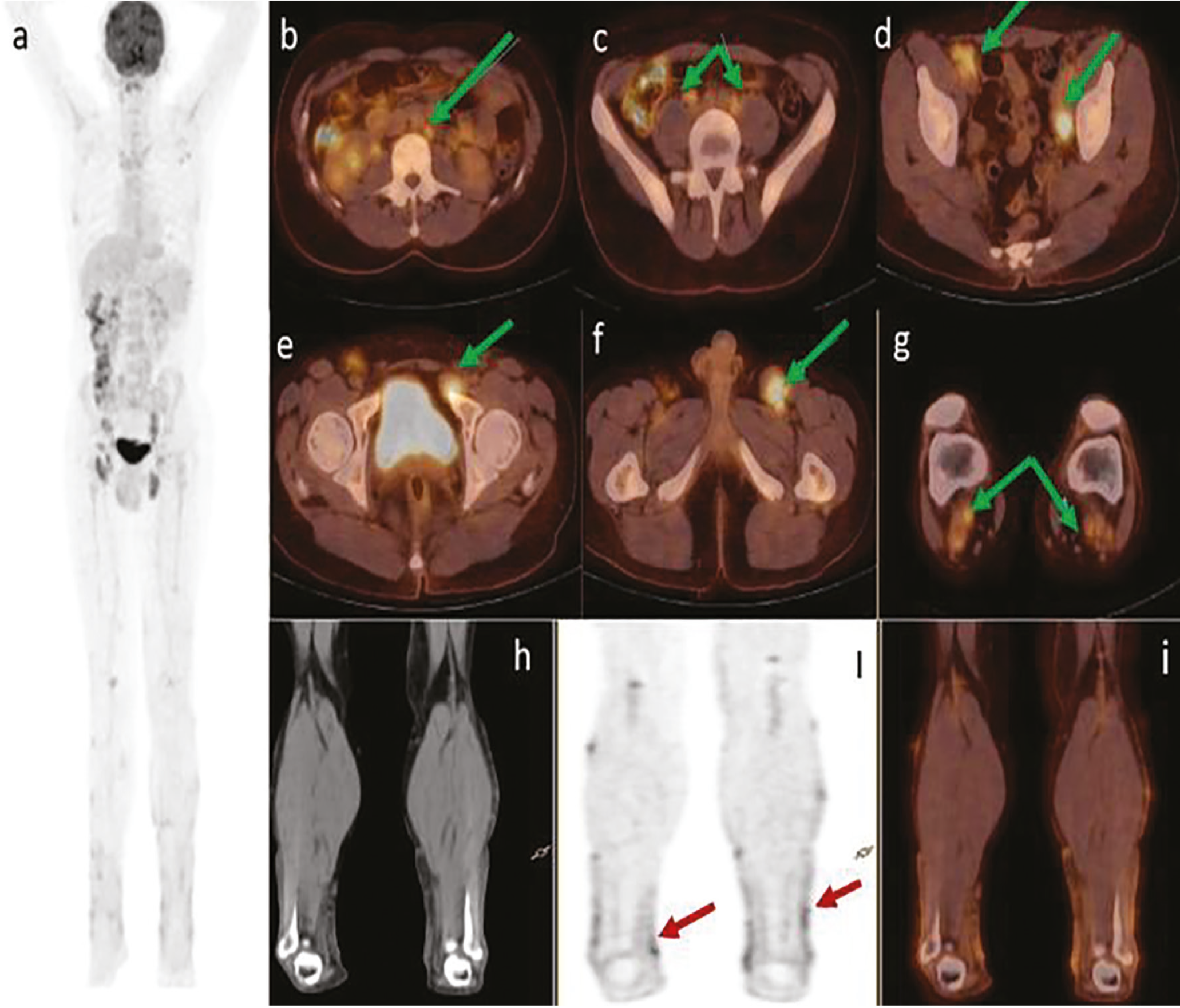
Figure 1
Figure 1. A 38-year-old male patient with weakness and fever. He had itchy skin lesions for 10 years and has been treated for dermatitis, but the lesions persisted despite treatment. Ultrasonography revealed multiple lymphadenopathies in both inguinal regions. In routine blood tests, C-reactive protein, white blood cells, sedimentation, urea, and blood urea nitrogen levels are high. Acute renal failure. Serum protein electrophoresis revealed hypoalbuminemia and hypergammaglobinemia. Human herpes virus 8 (HHV8) was negative. On physical examination, extensive erythematous excoriated plaques were observed on the trunk and extremities and purplish hyperkeratotic plaques with atrophic ulcerated areas in the middle were observed. Whole-body 18F-fluorodeoxyglucose positron emission tomography/computed tomography (18F-FDG PET/CT) maximum intensity projection images (a) and transaxial fusion PET/CT images revealed bilateral paraaortic (b), bilateral parailiac (c) (arrowhead; SUVmax: 3.5), bilateral external-internal iliac (d), left inguinal and femoral (e, f) (arrowhead; SUVmax: 6,3), bilateral popliteal (g) (arrowhead; SUVmax: 4.2) hypermetabolic lymph nodes (green arrows). Multiple hypermetabolic skin lesions are seen on transaxial CT (h), PET (ı; red arrows) and fusion PET/CT images (i).
View larger Download
Figure 2
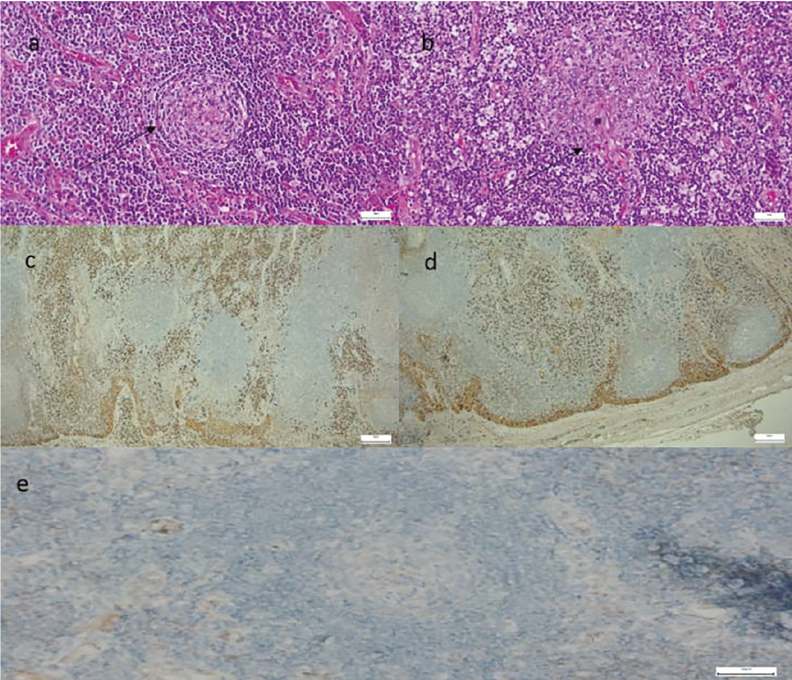
Figure 2. Biopsy of the left inguinal lymph node was performed. In microscopic examination, hyperplastic follicles were observed in the lymph nodes (a) (black arrow). In the hyperplastic follicles, the hyalinized vascular structures extended to the germinal center (b) (black arrow). Polyclonal staining was observed in plasma cells stained with kappa and lambda light chains (c, d). HHV8 was negative (e). Hodkin and non-Hodkin lymphoma were ruled out with Castleman’s disease 3 (CD) 3, CD 20, paxillin antigen expression in synovial tissue (PAXS), optical coherence tomography-2, B-cell lymphoma (Bcl)-2, Bcl-6, cyclin D1, CD4, CD8, CD30, epithelial membrane antigen, anaplastic lymphoma kinase, CD21, CD68, kappa, lambda, and Ki-67 antibodies. The pathological diagnosis was made as “CD, Plasma Cell Variant” with morphological and immunohistochemical findings. He was diagnosed with prurigo nodularis, lymphocytic vasculitis, and stasis dermatitis based on biopsies of skin lesions located in different regions. CD is a rare chronic lymphoproliferative disorder characterized by unexplained enlarged lymph nodes that was first described by Castleman et al. (1) in 1956. There are two clinical types of CD: unicentric CD and multicentric Castleman’s disease (MCD). MCD is a systemic disease characterized by multiple lymph nodes, in addition to symptoms such as fever, night sweats, and weight loss (2,3). Although rare, renal involvement can complicate the disease. Nephrotic syndrome, acute renal failure, interstitial nephritis, thrombotic microangiopathy, renal lymphoma, and renal amyloidosis have been reported (4). Skin involvement is very rare in CD. In the literature, skin lesions such as paraneoplastic pemphigus, lichenoid-nodular and maculopapular eruptions, Kaposi’s sarcoma, polyneuropathy, organomegaly, endocrinopathy, monoclonal gammopathy, skin changes syndrome, autoimmune bleeding disorders, cutaneous necrotizing vasculitis, and xanthogranuloma have been reported (5). A few case reports and one original article were found regarding the use of 18F-FDG-PET/CT in CD (5,6,7). In the study of Lee et al. (7), 4 of 12 patients with CD were unicentric, whereas 8 were multicentric. All lesions had moderate to high 18F-FDG uptake. There are no studies in the literature on 18F-FDG PET/CT in patients with MCD and skin lesions. Our case was clinically considered to be one of the skin diseases seen in CD patients. 18F-FDG draws attention as a case of acute renal failure with skin and multiple lymph node involvement on PET/CT. Studies on 18F-FDG PET/CT in MCD patients have been published. In a study by Jiang et al. (8), multiple lymphadenopathies as well as thickened skin with increased activity in the hip area were observed in a patient on 18F-FDG PET/CT images (SUVmax: 10.9). Skin biopsy revealed infection.
View larger Download

Footnotes

Ethics

Informed Consent: Patient consent was obtained.

Authorship Contributions

Surgical and Medical Practices: M.Y., T.Ş., Concept: T.Ş., Design: M.Y., T.Ş., N.Ş.T., Data Collection or Processing: N.Ş.T., Analysis or Interpretation: T.Ş., Literature Search: M.Y., Writing: M.Y.

Conflict of Interest: No conflicts of interest were declared by the authors.

Financial Disclosure: The authors declare that this study has received no financial support.

References

1

Castleman B, Iverson L, Menendez VP. Localized mediastinal lymphnode hyperplasia resembling thymoma. Cancer. 1956;9:822-830.

2

Cervantes CE, Correa R. Castleman disease: a rare condition with endocrine manifestations. Cureus. 2015;7:e380.

3

Kessler E. Multicentric giant lymph node hyperplasia. A report of seven cases. Cancer. 1985;56:2446-2451.

4

Uthup S, Balachandran K, Ammal VA, Abdul Salam R, George J, Nair GM, Leela M. Renal involvement in multicentric Castleman disease with glomeruloid hemangioma of skin and plasmacytoma. Am J Kidney Dis. 2006;48:e17-24.

5

Kim HJ, Han JH, Bang CH, Park KS, Cho SG, Yoo DS, Kim KM, Park HJ, Park YM, Lee JY, Lee JH. Cutaneous disorders associated with Castleman’s disease. Acta Derm Venereol. 2019;99:984-989.

6

Hwang JP, Kim J, Park JM. Human immunodeficiency virus negative multicentric Castleman’s disease coexistent with Kaposi’s sarcomaon 18F FDG PET/CT: A case report. Mol Clin Oncol 2019;10:318-320.

7

Lee ES, Paeng JC, Park CM. Metabolic characteristics of castleman disease on 18F-FDG PET in relation to clinical Implication. Clin Nucl Med 2013;38:339-342.

8

Jiang Y, Hou G, Zhu Z, Huo L, Li F, Cheng W. 18F-FDG PET/CT imaging features of patients with multicentric Castleman disease. Nucl Med Commun. 2021;42:833-838.