

Abstract
Prostatic adenocarcinoma is characterized by elevated phosphatidylcholine metabolism. 18F-choline positron emission tomography/computed tomography (PET/CT) is widely used for patients with biochemical recurrence and a prostate-specific antigen threshold above 2 ng/mL. We report a case of a patient with high-risk prostatic adenocarcinoma undergoing 18F-choline PET/CT for biochemical recurrence. In addition to hypermetabolic abdominal lymph nodes, an unexpected right testicular hypermetabolism was observed. Such findings on 18F-choline PET/CT may suggest a primary tumor or testicular metastasis of prostate cancer. Bilateral orchiectomy revealed a vitelline tumor associated with known primary prostatic cancer. The incidental discovery of a testicular vitelline tumor during prostate cancer imaging is rare, highlighting the importance of thorough diagnostics. This case underscores the need for comprehensive care in managing complex and atypical cancer cases, emphasizing the potential for unrelated tumor discoveries during diagnostic workup. Further research is essential for a better understanding of these rare cooccurring cancers and their treatment implications.
Keywords: 18F-choline positron emission tomography/computed tomography, yolk tumor, prostatic adenocarcinoma
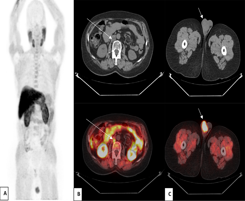
In the CT and fusion images in axial sections (arrow; C), we also noted the presence of a suspicious right testicular nodule measuring 43x30 mm with an intense hypermetabolism (SUVmax =8.8). Our findings suggest two diagnoses: primary cancer or rare testicular metastasis of prostate cancer.
Rare testicular hypermetabolism requires prompt reporting by the nuclear medicine physician, prompting a follow-up testicular ultrasound, especially if CT scan shows suspicious abnormalities. Testicular ultrasonography showed two contiguous, well-limited, heterogeneous tissue nodules taking color Doppler and containing microcalcifications, measuring 23x27 mm and 19x20 mm respectively.
Few cases of testicular involvement in prostate cancer have been reported in the literature using 18F-choline PET/CT. Identifying a testicular lesion in a familial neoplastic context, particularly in prostatic cases, requires careful investigation and histological confirmation to distinguish metastatic from primary origin (1).
Moreover, the presence of this unusual testicular hypermetabolism poses challenges in terms of differential diagnosis, especially testicular secondary involvement, which is uncommon (2-3% prevalence) and is typically discovered during autopsy or orchidectomy. Prostate cancer nearly constitutes half of all testicular metastases, with suggested dissemination through different ducts or vascular networks, emphasizing an unconventional metastatic pathway. Foret et al. (5) reported a rare instance of isolated testicular recurrence of prostatic adenocarcinoma identified via 18F-fluorocholine PET/CT. Furthermore, there are no studies in the literature addressing the various diagnoses to consider in the presence of this testicular hypermetabolism, particularly benign testicular pathologies unrelated to neoplastic contexts.
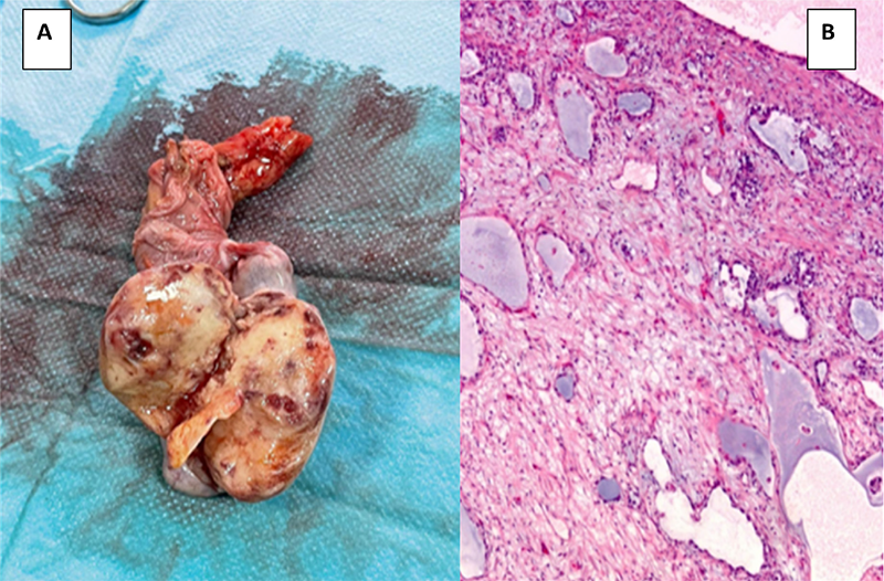
This case underscores the pivotal role of 18F-choline PET/CT in the management of prostatic neoplasia, revealing a rare primary testicular cancer. It highlights the need for additional scientific studies and emphasizes the importance of systematic pathological examination post-surgical castration for improved differential diagnosis and enhanced control of prostatic neoplasia, especially after addressing lymph node recurrence. In addition, the discovery of a testicular vitelline tumor during 18F-choline investigation for prostate cancer emphasizes the significance of thorough imaging and testing. It stresses the requirement for comprehensive, multidisciplinary care in addressing complex cases, urging further research to understand the implications and potential treatment strategies for these uncommon concurrent cancers.
Footnotes
Ethics
Informed Consent: Informed consents of the patient was obtained.
Authorship Contributions
Concept: C.B., S.O.N., M.A., O.A.S., Y.B., A.D., Design: C.B., S.O.N., O.A.S., Y.B., A.D., Data Collection or Processing: C.B., Analysis or Interpretation: C.B., S.O.N., A.D., Literature Search: C.B., M.A., Writing: C.B.
Conflict of Interest: No conflicts of interest were declared by the authors.
Financial Disclosure: The authors declare that this study has received no financial support.
References
Bauduceau O, Vedrine L, Chargari C, Ceccaldi B, Le Moulec S, Houlgatte A. Métastase testiculaire d’un adénocarcinome prostatique: à propos d’un cas [Testicular metastasis of prostatic adenocarcinoma: a case report]. Prog Urol. 2007;17:251-252. French.
Zizi M, Ziouziou I, El Yacoubi S, Khmou M, Jahid A, Mahassini N, Karmouni T, El Khader K, Koutani A, Andaloussi AI. Tumeur du sac vitellin du testicule au stade IIIc métastatique : à propos d’un cas. Can Urol Assoc J. 2014;8:E663-E665. French.
Calabrò A, Bosio G, Drera E, Giubbini R, Bertagna F. Incidental Finding of Testicular Seminoma by 18F-Choline PET/CT in a Prostate Cancer Patient. Clin Nucl Med. 2022;47:e249-e251.
Cegla P, Scibisz-Dziedzic K, Witkowska K, Kubiak A, Wierzchoslawska E, Kycler W, Chrapko B, Czepczyński R. Detection of a second primary cancer in a 18F-fluorocholine PET/CT - multicentre retrospective analysis on a group of 1345 prostate cancer patients. Nucl Med Rev Cent East Eur. 2022;25:25-30.
Foret T, Dimier JP, Skanjeti A, Dhomps A, Tordo J. Récidive testiculaire isolée d’un adénocarcinome prostatique révélée par TEP/TDM 18F-fluorocholine [Solitary testicular metastasis from prostate adenocarcinoma revealed byF-fluorocholine PET/CT]. Médecine Nucléaire. 2020;44:76-78.

