

Abstract
Primary peritoneal serous carcinoma (PPSC) is a rare malignancy that mostly affects women. In males, there are only few reports in the literature. 18F-fluorodeoxyglucose (18F-FDG) positron emission tomography/computed tomography (PET/CT) is useful for evaluating the origin of the tumor, its extent, and distant metastasis. Moreover, 18F-FDG PET/CT was helpful in distinguishing between PPSC and peritoneal carcinomatosis. We present a case of PPSC on 18F-FDG PET/CT in a male with histopathological correlation.
Keywords: Peritoneal tumour, primary peritoneal serous carcinoma, 18F-FDG PET/CT
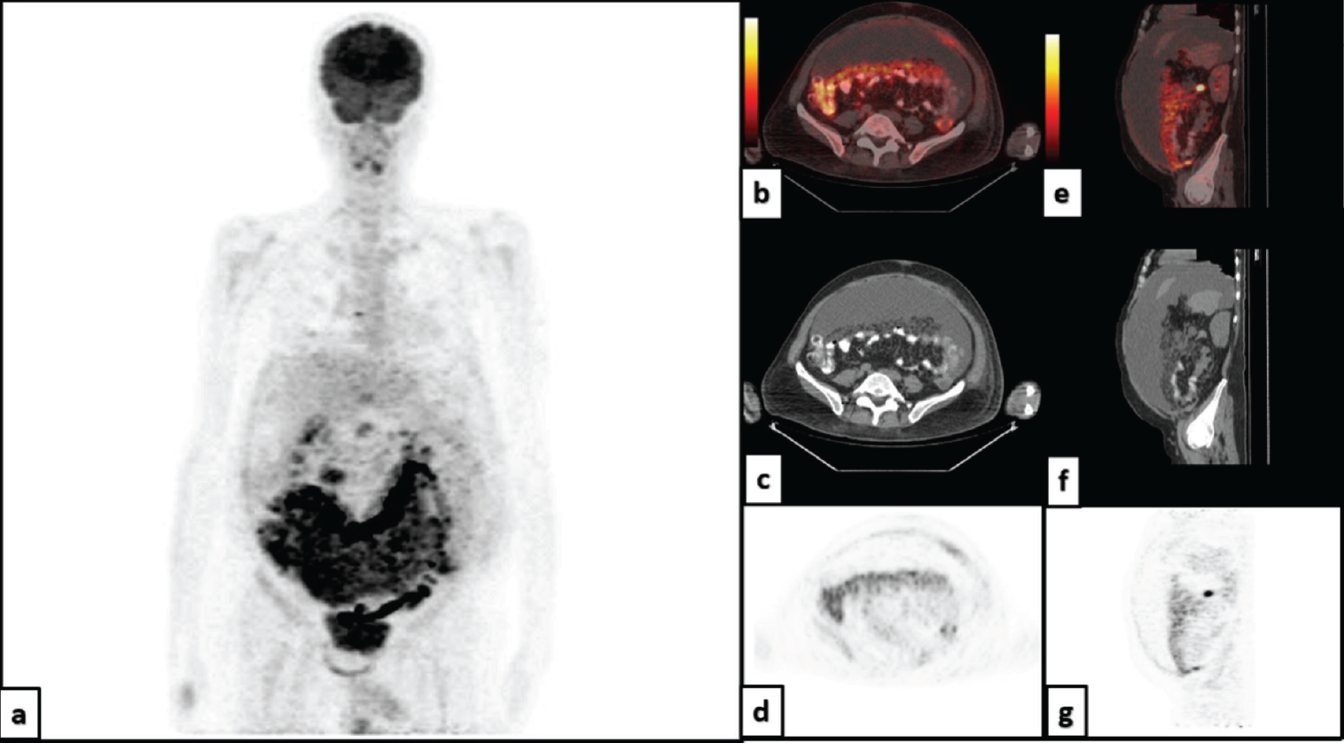
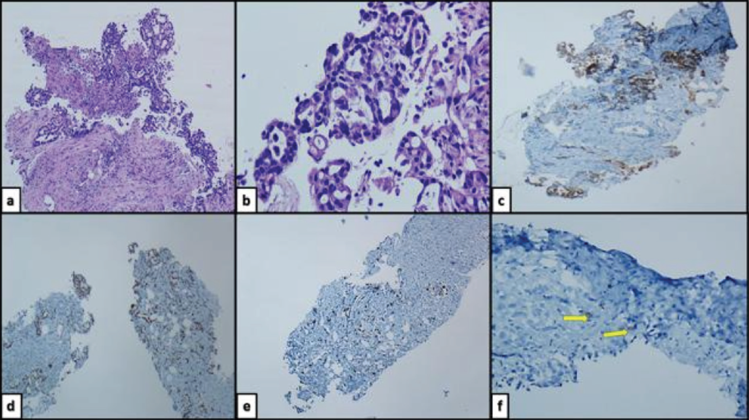
Primary peritoneal serous carcinoma (PPSC) is a rare epithelial tumor that occurs almost exclusively in women (1,2). The male: female ratio ranged from 0.0018 to 0.0045 (3). There are only a few case reports of PPSC developing in men (4). Histopathological and cytological characteristics of these tumors are similar to those of epithelial ovarian cancer (2). Therefore, in addition to histopathological examination and imaging, ovarian cancer must be ruled out during diagnosis. Imaging methods such as CT, MRI, and PET/CT are used to confirm diagnosis, differentiate between primary peritoneal cancer and peritoneal metastasis, and determine the spread of the disease (5). Although not sufficient for diagnosis, 18F-FDG PET/CT can assist in distinguishing between PPSC and peritoneal karsinomatosis (6). 18F-FDG PET/CT is useful in evaluating the origin of the tumor, its extent, and distant metastasis (3).
Footnotes
Ethics
Informed Consent: Patient consent was obtained.
Authorship Contributions
Surgical and Medical Practices: B.Ö.G., B.G., F.Ü., Concept: N.C., F.Ü., Design: B.G., Data Collection or Processing: B.Ö.G., Analysis or Interpretation: B.G., N.C., Literature Search: B.Ö.G., Writing: B.Ö.G., F.Ü.
Conflict of Interest: No conflicts of interest were declared by the authors.
Financial Disclosure: The authors declare that this study has received no financial support.
References
Neuhausen SL, Shani H, Boker LK, Steele L, Silverman BG, Ottini L, Silvestri V, Laitman Y, Korach J, Perri T, Friedman E. Primary peritoneal serous carcinoma in men: A rare and non-BRCA-associated entity. Anticancer Res. 2017;37:3069-3072.
Levy AD, Arnáiz J, Shaw JC, Sobin LH. From the archives of the AFIP: primary peritoneal tumors: imaging features with pathologic correlation. Radiographics. 2008;28:583-607.
Guellil A, Jabi R, Mabrouk MY, Bouzayan L, Merhoum A, Del Gallo G, Godart C, Bouziane M. Primary peritoneal high-grade serous carcinoma in a man: A case report. Ann Med Surg (Lond). 2022;77:103605.
Gan J, Herzog J, Smith DA, Vos D, Kikano E, Tirumani SH, Ramaiya NH. Primary peritoneal serous carcinoma: a primer for radiologists. Clin Imaging. 2022;83:56-64.
Turlakow A, Yeung HW, Salmon AS, Macapinlac HA, Larson SM. Peritoneal carcinomatosis: role of (18)F-FDG PET. J Nucl Med. 2003;44:1407-1412.
Yun WS, Bae JM. Primary peritoneal serous carcinoma, an extremely rare malignancy: A case report and review of the literature. Oncol Lett. 2016;11:4063-4065.

