

Abstract
The uptake of Tc-99m-uptake of Tc-99m-methylene diphosphonate (MDP) on bone scintigraphy can be seen at sites other than bone in a varying number of benign and malignant conditions. Extraosseous metastatic calcifications can occur in ovarian papillary serous adenocarcinoma (PSAC). These extraosseous calcifications show Tc-99m-MDP uptake. We report a case of a female in her sixties who had a previous history of PSAC of the ovary. The patient had undergone neoadjuvant chemotherapy (NACT) followed by total abdominal hysterectomy and bilateral salpingo-oophorectomy. She also received adjuvant chemotherapy. Tc-99m-MDP bone scan was performed post chemotherapy because the patient complained of lower backache. The scan showed increased uptake in the lower thoracic and lumbar vertebral regions. However, single-photon emission computed tomography/computed tomography (CT) localizes the uptake to metastatic calcified peritoneal deposits. Further 18F-fluorodeoxyglucose positron emission tomography/CT confirmed widespread peritoneal and omental metastatic disease with increased uptake.
Keywords: Tc-99m-MDP, metastases, ovarian papillary serous adenocarcinoma
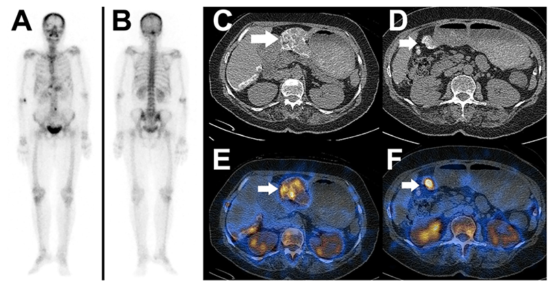
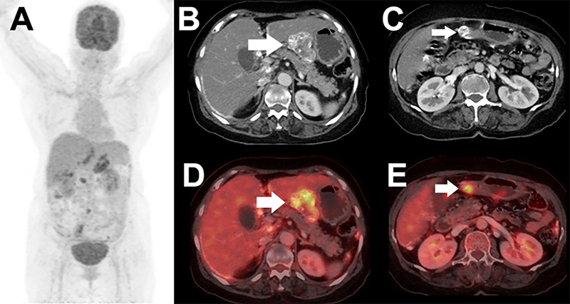
Many previously published studies have shown the uptake of Tc-99m-MDP in calcified metastases from Krukenberg tumor (4), endometrium, and ovary (5,6). This uncommon extraosseous accumulation can be explained by the affinity of Tc-99m-MDP for hydroxyapatite crystals and calcium precipitates (7) in soft tissue metastases from ovarian PSAC. Another explanation could be altered extracellular fluid and tracer handling dynamics and tumor neovascularization with altered capillary permeability (4,8). This extraosseous uptake on bone scans leads to misdiagnosis of bone metastases. Hybrid imaging like SPECT/CT, provides additional anatomic information and leads to localization of tracer uptake for further clarification, as in this case.
Footnotes
Ethics
Informed Consent: Written informed consent has been obtained from the patient for the publication of the case.
Authorship Contributions
Concept: K.A., P.S.S.P., G.K.P., Data Collection or Processing: K.B., Analysis or Interpretation: K.B., K.A., P.S.S.P., G.K.P., Literature Search: K.B., Writing: K.B., K.A.
Conflict of Interest: No conflicts of interest were declared by the authors.
Financial Disclosure: The authors declare that this study has received no financial support.
References
Momenimovahed Z, Tiznobaik A, Taheri S, Salehiniya H. Ovarian cancer in the world: epidemiology and risk factors. Int J Womens Health. 2019;11:287-299.
Bharwani N, Reznek RH, Rockall AG. Ovarian Cancer Management: the role of imaging and diagnostic challenges. Eur J Radiol. 2011;78:41-51.
Sharma SK, Nemieboka B, Sala E, Lewis JS, Zeglis BM. Molecular Imaging of Ovarian Cancer. J Nucl Med. 2016;57:827-833.
Oppenheimer AG, Onyedika C, Freeman LM. MDP uptake in peritoneal carcinomatosis and Krukenberg tumors from mucinous adenocarcinoma. Clin Nucl Med. 2011;36:235-236.
Radin AI, Youssef IM, Quimbo RD, Perone RW, Guerrieri C, Abdel-Dayem HM. Technetium-99m diphosphonate imaging of psammocarcinoma of probable ovarian origin: case report and literature review. Clin Nucl Med. 2005;30:395-399.
Ozulker T, Ozulker F, Ozpacaci T. 99mTc-MDP and 18F-FDG uptake in calcified metastatic lesions from ovarian papillary serous adenocarcinoma. Hell J Nucl Med. 2009;12:287-288.
Kaye J, Hayward M. Soft tissue uptake on 99mTc methylene diphosphonate bone scan imaging: pictorial review. Australas Radiol. 2002;46:13-21.
Agrawal K, Bhattacharya A, Harisankar CN, Abrar ML, Dhaliwal LK, Mittal BR. F-18 fluoride uptake in calcified extraosseous metastases from ovarian papillary serous adenocarcinoma. Clin Nucl Med. 2012;37:e22-e23.

