

Abstract
Bone marrow necrosis (BMN) is usually associated with malignancies and is characterized by multiple geographic signal abnormalities on magnetic resonance imaging (MRI). We report a 28-year-old female with BMN and underlying diffuse large B-cell lymphoma. Diffuse abnormal signal intensities through the vertebral column were demonstrated on her pretreatment MRI, and the diagnosis of BMN was challenging. Positron emission tomography/computed tomography (PET/CT) for lymphoma staging showed multiple decreased or absent 18F-fluorodeoxyglucose (18F-FDG) uptake within the vertebrae and pelvis. Marrow biopsy pathological examination showed lymphoma infiltration and massive necrosis. On the follow-up MRI obtained approximately 21 months after the PET/CT scan, multiple geographic abnormal signal intensities were detected within the vertebral column and were consistent with the areas of decreased 18F-FDG uptake on PET/CT. This case indicates that 18F-FDG PET/CT is helpful in the diagnosis of BMN with atypical MRI appearances.
Keywords: Bone marrow necrosis, lymphoma, 18F-FDG PET/CT, MRI
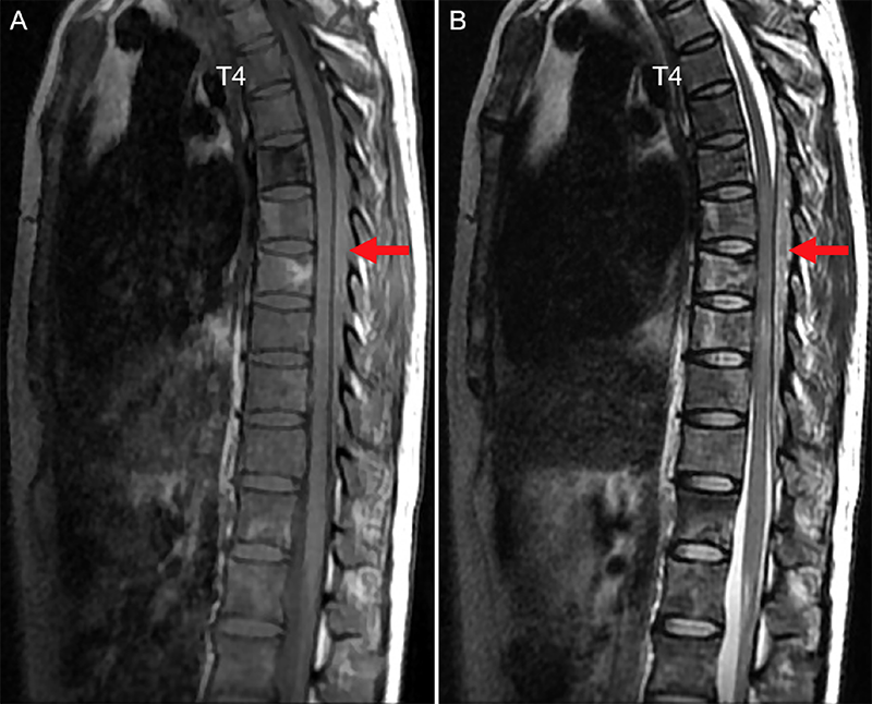
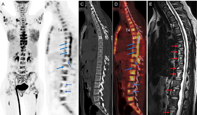
BMN is caused by microcirculation failure (1) and is characterized as necrosis of myeloid tissue and medullary stroma. BMN is usually associated with malignancies and mainly involves the spine and pelvis (1). It can lead to bone pain, fever, and fatigue (2). Sulfur colloid marrow scan was once thought to be useful in the diagnosis and assessment of the extent of BMN (3,4,5). Later, BMN was more commonly evaluated by MRI with its characteristic geographic signal abnormalities: central hyperintensity without enhancement surrounded by a peripheral band of hypointense signal (6,7). However, the diagnosis of BMN may be challenging based on MRI because MRI appearances may vary at different stages of BMN and may be influenced by the underlying marrow composition. As shown in this study, typical geographic signal abnormalities were not obvious on preoperative MRI. However, BMN should be suspected based on PET/CT findings of decreased or absent 18F-FDG uptake in multiple vertebrae (8). The case indicates that PET/CT is complementary to MRI in the early diagnosis of BMN.
Footnotes
Ethics
Informed Consent: The written informed consent has been obtained from the patient.
Authorship Contributions
Surgical and Medical Practices: L.S., Concept: W.Z., Design: W.Z., Data Collection or Processing: L.S., H.L., Analysis or Interpretation: L.S., W.Z., Literature Search: L.S., H.L., Writing: L.S.
Conflict of Interest: No conflicts of interest were declared by the authors.
Financial Disclosure: The authors declare that this study has received no financial support.
References
Janssens AM, Offner FC, Van Hove WZ. Bone marrow necrosis. Cancer. 2000;88:1769-1780.
Murphy PT, Sivakumaran M, Casey MC, Liddicoat A, Wood JK. Lymphoma associated bone marrow necrosis with raised anticardiolipin antibody. J Clin Pathol. 1998;51:407-409.
Carloss H, Winslow D, Kastan L, Yam LT. Bone marrow necrosis. Diagnosis and assessment of extent of involvement by radioisotope studies. Arch Intern Med. 1977;137:863-866.
Navari RM, Carter J, Hillman RS. Bone marrow necrosis in acute leukemia. Acta Haematol. 1983;69:158-163.
Cowan JD, Rubin RN, Kies MS, Cerezo L. Bone marrow necrosis. Cancer. 1980;46:2168-2171.
Weissman DE, Negendank WG, al-Katib AM, Smith MR. Bone marrow necrosis in lymphoma studied by magnetic resonance imaging. Am J Hematol. 1992;40:42-46.
Tang YM, Jeavons S, Stuckey S, Middleton H, Gill D. MRI features of bone marrow necrosis. AJR Am J. Roentgenol. 2007;188:509-514.
Dong P, Tian R, Li L, Su M. Bone marrow necrosis secondary to metastatic adenocarcinoma revealed by 18F-FDG PET/CT: A clinical case report. Medicine (Baltimore). 2017;96:e9067.

