Unexpected Detection of Cephalad Renal Ectopia Due to Large Omphalocele Containing the Liver on Tc-99m DMSA Scintigraphy
PDF

Interesting Image

Unexpected Detection of Cephalad Renal Ectopia Due to Large Omphalocele Containing the Liver on Tc-99m DMSA Scintigraphy

Mol Imaging Radionucl Ther 2025;34(1):85-87 DOI: 10.4274/mirt.galenos.2024.68095
, ,
PDF
Permissions

Abstract

Omphalocele is a congenital abdominal wall defect with herniation of abdominal viscera into a sac. Tc-99m DMSA renal cortical scan is a functional imaging technique used for detecting parenchymal defects, mostly in patients with recurrent urinary tract infection as well as congenital renal abnormalities. Renal anomalies are known to accompany omphalocele. In this retrospective study, we present a case of cephalad renal ectopia as observed on Tc-99m DMSA scintigraphy in a patient with omphalocele due to a large hernia sac containing most of the liver; and we review the renal abnormalities associated with omphalocele in the literature.

Keywords: Omphalocele, cephalad renal ectopia, Tc-99m DMSA, liver, renal abnormality
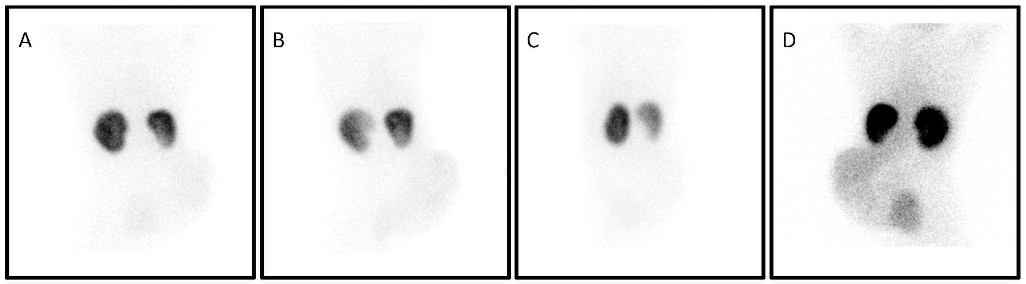
Figure 1
Figure 1. A 6-year-old girl with omphalocele was referred to our clinic with the diagnosis of recurrent urinary tract infection (UTI) after a Tc-99m DMSA scan. The Tc-99m DMSA scan revealed minimally decreased Tc-99m DMSA uptake in the lower pole of the right kidney, which may be suggestive of a parenchymal defect due to UTI and otherwise quite normal Tc-99m DMSA biodistribution throughout both kidneys. However, the kidneys appeared to be located more cranially than their normal expected position within the abdomen, along with an impression suspicious of a rotational anomaly seen on posterior, right posterior oblique, and left posterior oblique projections (Figure 1A, B, C) respectively. To our knowledge, there was also an unusual Tc-99m DMSA uptake in the right abdominal quadrant, with a laterally bulging appearance. The pattern of the uptake was homogeneous, and its intensity was lower than that of Tc-99m DMSA uptake in the kidneys, and the intensity of such uptake was more prominent on the anterior projection image (Figure 1D). Thus, these findings suggest that the uptake may not be due to an ectopic kidney as a component of supernumerary kidneys. Upon physical examination, a large hernia sac with an omphalocele was observed in the area.
View larger Download

Figure 2
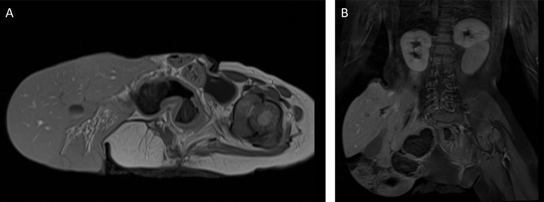
Figure 2. Magnetic resonance imaging (Figures 2a axial, 2b coronal) revealed a large omphalocele that included most of the liver, corresponding to the faint but homogeneous Tc-99m DMSA uptake in the right quadrant of the abdomen (Figure 2a). The kidneys were located just below the diaphragm, consistent with cephalad renal ectopia, and the liver was mostly located within the omphalocele sac (Figure 2b). Omphalocele is an abdominal wall defect characterized by herniation of the abdominal viscera into a sac (1). During fetal development, omphalocele occurs when gut contents fail to rotate and return to the abdominal cavity (2). It is a rare congenital defect that shows a severity spectrum from small umbilical hernia to large sac with evisceration of all abdominal organs, as in our case (3). Omphalocele is frequently associated with syndromes and a variety of additional congenital abnormalities, including chromosomal abnormalities, cardiac, pulmonary, gastrointestinal, musculoskeletal manifestations, and neural tube defects (3,4,5,6). In addition, associated renal anomalies can be observed. The kidneys migrate from the pelvis to the upper abdomen during normal fetal development. The liver plays an important role in the normal position of the kidneys as it arrests the ascent of the kidneys. Omphalocele containing the liver has been accounted for excessive migration, which is finally stopped by the diaphragm, resulting in cephalad renal ectopia (7,8). Our patient had a neural tube defect, right lower extremity agenesia, cephalad renal ectopia, and omphalocele. To the best of our knowledge, this is the first case of a laterally protruded large omphalocele containing most of the liver that was diagnosed with cephalad renal ectopia on a Tc-99m DMSA scan.
View larger Download

Footnotes

Ethics

Informed Consent: An informed consent was obtained from the patient.

Authorship Contributions

Surgical and Medical Practices: Z.I., C.K., Concept: Z.I., M.F.B., Design: Z.I., M.F.B., Data Collection or Processing: Z.I., C.K., Analysis or Interpretation: Z.I., M.F.B., Literature Search: Z.I., C.K., M.F.B., Writing: Z.I., C.K., M.F.B.

Conflict of Interest: No conflicts of interest were declared by the authors.

Financial Disclosure: The authors declare that this study has received no financial support.

References

1

Fernbach SK. Neonatal Gastrointestinal Radiology. In:Textbook of Gastrointestinal Radiology W.B. Saunders; 2008:2203-2233.

2

Zahouani T, Mendez MD. Omphalocele. In: StatPearls [Internet]. Treasure Island (FL): StatPearls. 2021 Jan–.

3

Gaca M, Bissett GS. Diseases of the Pediatric Abdominal Wall, Peritoneum, and Mesentery. In: Textbook of Gastrointestinal Radiology. W.B. Saunders. 2008: 2371-2381.

4

Bence CM, Wagner AJ. Abdominal wall defects. Transl Pediatr. 2021;10:1461-1469.

5

Conner P, Vejde JH, Burgos CM. Accuracy and impact of prenatal diagnosis in infants with omphalocele. Pediatr Surg Int. 2018;34:629-633.

6

Corey KM, Hornik CP, Laughon MM, et al. Frequency of anomalies and hospital outcomes in infants with gastroschisis and omphalocele. Early Hum Dev. 2014;90:421-424.

7

Connolly LP, Connolly SA, Treves ST. Renal malposition secondary to omphalocele shown on hepatobiliary scintigraphy. Clin Nucl Med. 2001;26:546-548.

8

Pinckney LE, Moskowitz PS, Lebowitz RL, et al. Renal malposition associated with omphalocele. Radiology. 1978;129:677-682.