

Abstract
Intrathyroidal parathyroid adenoma is a rare anomaly with an incidence of 1.4-6%. A cause of failure in the therapeutic management of hyperparathyroidism is the ectopic localization of the adenoma, particularly intrathyroidal. This is the case of a 51-year-old patient with no particular pathological history who presented with neck pain and whose clinical examination revealed a goiter. Parathyroid myocardial perfusion imaging (MIBI) scintigraphy revealed an elective MIBI-fixing focus with an upper left polar projection compatible with a parathyroid origin. A left loboishmectomy was performed. Anatomopathological study showed a parathyroid adenoma associated with dystrophic thyroid parenchyma, with no obvious histological signs of malignancy. Post-therapeutic laboratory work-up was normalized. Parathyroid adenoma, intrathyroidal, is an uncommon lesion but may be responsible for therapeutic failure in hyperparathyroidism. MIBI parathyroid gland scintigraphy is the gold standard for the diagnosis of parathyroid ectopy.
Keywords: Intrathyroidal parathyroid adenoma, parathyroid ectopy, MIBI scintigraphy
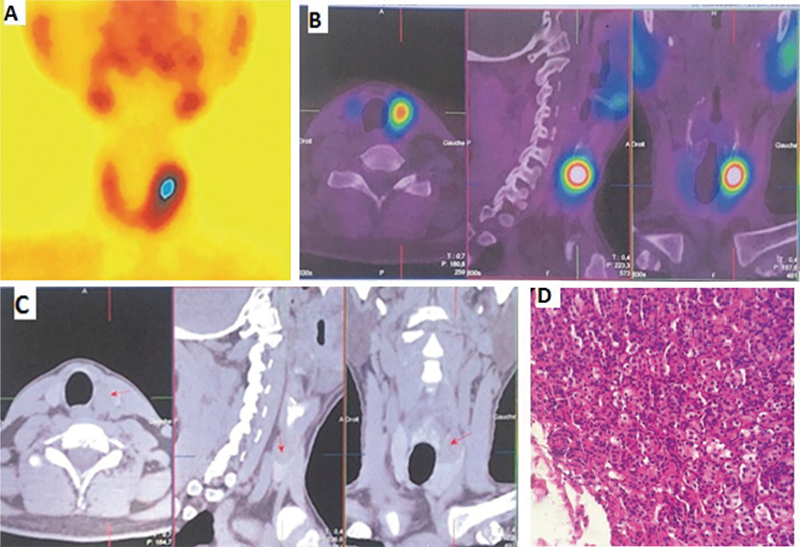
Hyperparathyroidism is the third most common endocrinopathy, with parathyroid adenoma being the most common etiology. The latter may be located ectopically in 11-25% of cases, secondary to embryonic migration anomalies (1). Intrathyroidal parathyroid adenoma is a rare anomaly with an incidence of 1.4-6%. One of the causes of failure in the therapeutic management of hyperparathyroidism is the ectopic localization of the adenoma, particularly intrathyroidal (2). The diagnosis of the location of the ectopic gland depends mainly on imaging studies, such as ultrasound, CT, magnetic resonance imaging (MRI), Tc-99m-MIBI SPECT/CT, or 18F-fluorocholine positron emission tomography (PET)/CT (3). Tc-99m-MIBI SPECT/CT represents additional advantages in the evaluation of parathyroid adenoma. It can be used to estimate the function of the parathyroid gland and the extent of its hyperplasia is the simplest and most widely used noninvasive method for preoperative determination of the location and position of the gland (4). Numerous studies have confirmed the usefulness of MIBI scintigraphy in the exploration of the parathyroid gland in the management of hyperparathyroidism. In a group of over 1,500 cases, Roy et al. (5) observed a parathyroidectomy success rate of over 95% and the presence of an ectopic parathyroid gland in 6-16% of cases (38% located in the thymus, 31% in the retropharynx and 18% intra-thyroid). In such cases, these authors found MIBI scintigraphy was diagnostically superior, with a sensitivity of 89% compared with ultrasound (sensitivity 59%) (5). Ishibashi et al. (6) recommended that MIBI scintigraphy be performed before any other diagnostic means of localization, such as CT or MRI. This author observed a sensitivity of 70% and a specificity of 88% for scintigraphy, which is superior to CT (40% and 88% respectively) and MRI (60% and 88%) (6). Other studies also point to MIBI scintigraphy as the best performing diagnostic technique, with sensitivity approaching 90% (7). However, MIBI scintigraphy has certain limitations. For example, when parathyroid hyperplasia is not evident, or the focus is too small to be covered by thyroid tissue, or when associated with a multinodular goiter, false-negative results can occur; hence, it is also of interest in the differential diagnosis of benign thyroid disease. Furthermore, when ultrasound and Tc-99m-MIBI SPECT/CT cannot determine intrathyroid nodules, PET/CT may be a better alternative (8).
Footnotes
Ethics
Informed Consent: The patient consent was obtained.
Authorship Contributions
Surgical and Medical Practices: S.Z., M.A.B., A.M., A.Mar., I.Z.T., Concept: S.Z., Design: S.Z., Data Collection or Processing: S.Z., Analysis or Interpretation: S.Z., M.A.B., A.M., A.Mar., I.Z.T., Literature Search: S.Z., Writing: S.Z.
Conflict of Interest: No conflicts of interest were declared by the authors.
Financial Disclosure: The authors declare that this study has received no financial support.
References
Kaplan EL, Yashiro T, Salti G. Primary hyperparathyroidism in the 1990s. Choice of surgical procedures for this disease. Ann Surg. 1992;215:300-317.
Mazeh H, Kouniavsky G, Schneider DF, Makris KI, Sippel RS, Dackiw AP, Chen H, Zeiger MA. Intrathyroidal parathyroid glands: small, but mighty (a Napoleon phenomenon). Surgery. 2012;152:1193-1200.
Hai R, Xie LJ, You Q, Wu F, Qiu GC, Zhou XY. Diagnosis of Ectopic Intrathyroidal Parathyroid Adenoma with Nodular Goiter by 18F Fluorocholine: A Case Report. Ear Nose Throat J. 2022:1455613221103082.
Özdemir E, Genç M, Aydos U, Polat ŞB, Kandemir Z, Tam AA, Yildirim N, Türkölmez Ş. Comparison of 99mTc-MIBI planar scintigraphy, SPET/CT and ultrasonography in detection of parathyroid adenoma in patients with primary hyperparathyroidism. Hell J Nucl Med. 2020;23:21-26.
Roy M, Mazeh H, Chen H, Sippel RS. Incidence and localization of ectopic parathyroid adenomas in previously unexplored patients. World J Surg. 2013;37:102-106.
Ishibashi M, Nishida H, Hiromatsu Y, Kojima K, Uchida M, Hayabuchi N. Localization of ectopic parathyroid glands using technetium-99m sestamibi imaging: comparison with magnetic resonance and computed tomographic imaging. Eur J Nucl Med. 1997;24:197-201.
Castellani M, Reschini E, Longari V, Paracchi A, Corbetta S, Marotta G, Gerundini P. Role of Tc-99m sestamibi scintigraphy in the diagnosis and surgical decision-making process in primary hyperparathyroid disease. Clin Nucl Med. 2001;26:139-144.
Piccardo A, Trimboli P, Puntoni M, Foppiani L, Treglia G, Naseri M, Bottoni GL, Massollo M, Sola S, Ferrarazzo G, Bruzzone M, Catrambone U, Arlandini A, Paone G, Ceriani L, Cabria M, Giovanella L. Role of 18F-Choline Positron Emission Tomography/Computed Tomography to Detect Structural Relapse in High-Risk Differentiated Thyroid Cancer Patients Thyroid. 2019;29:549-556.

