

Abstract
A man in his 30s awaiting end-of-treatment 18F-fluorodeoxyglucose positron emission tomography/CT (18F-FDG PET/CT) for primary mediastinal B-cell lymphoma developed chest pain and pericardial effusion. His interim 18F-FDG PET/CT showed complete metabolic responses. His blood test revealed elevated levels of inflammatory markers, including C-reactive protein of 204.1 mg/L and erythrocyte sedimentation rate of 106 mm/h. His pericardial biopsy revealed organizing fibrinous pericarditis with hemosiderin pigment deposition and no evidence of malignancy or granuloma. The 18F-FDG PET/CT performed during this episode of illness revealed a mild degree of 18F-FDG uptake along the pericardial lining [maximum standardized uptake value (SUVmax) =6.76] compared with the blood pool activity (SUVmax =3.17), which favors pericarditis over relapsed lymphoma. His symptoms subsided 2 weeks after treatment with an non-steroidal anti-inflammatory drug, and he had no sign of relapsed lymphoma on subsequent follow-ups.
Keywords: Nuclear medicine, pericarditis, non-Hodgkins lymphoma, cardiology
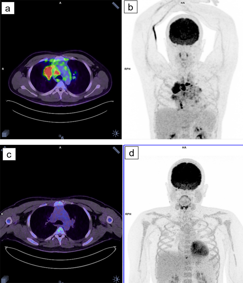
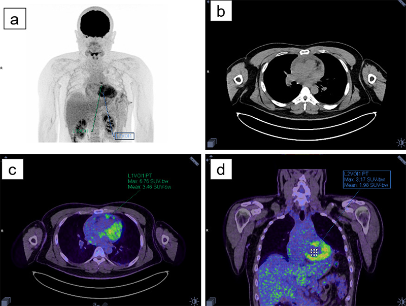
Although 18F-FDG PET/CT has not yet been established as a standard investigation for pericarditis, potential uses of 18F-FDG PET/CT have been proposed for multiple pericardial diseases (1). One potential use of 18F-FDG PET/CT is to differentiate between malignant and benign pericardial diseases. A study by Shao et al. (2) proposed a cut-off of SUVmax lesion/SUVmax blood pool ratio of >2.4 to indicate malignant pericardial disease with a sensitivity of 92.3% and specificity of 90.0%. In this study, the SUVmax ratio was 2.13, which correctly classified the uptake as benign. Other potential uses of 18F-FDG PET/CT in acute idiopathic pericarditis include predicting the risk of pericarditis relapse (3) and differentiating tuberculous from idiopathic pericarditis (4,5). Our case report supports the idea that 18F-FDG PET/CT can be a reliable diagnostic tool for differentiating malignant from non-malignant pericardial diseases.
Footnotes
Ethics
Informed Consent: The Institutional Review Board of the Faculty of Medicine at Chulalongkorn University approved this case and waived the requirement for written informed consent based on its retrospective nature (COE No. 020/2023).
Authorship Contributions
Surgical and Medical Practices: C.B., Concept: N.B., Design: N.B., Data Collection or Processing: C.B., Analysis or Interpretation: S.V., N.B., Literature Search: S.V., N.B., Writing: S.V., N.B.
Conflict of Interest: No conflicts of interest were declared by the authors.
Financial Disclosure: The authors declare that this study has received no financial support.
References
Kim MS, Kim EK, Choi JY, Oh JK, Chang SA. Clinical utility of [18F]FDG-PET /CT in pericardial disease. Curr Cardiol Rep. 2019;21:107.
Shao D, Wang SX, Liang CH, Gao Q. Differentiation of malignant from benign heart and pericardial lesions using positron emission tomography and computed tomography. J Nucl Cardiol. 2011;18:668-677.
Gerardin C, Mageau A, Benali K, Jouan F, Ducrocq G, Alexandra JF, van Gysel D, Papo T, Sacre K. Increased FDG-PET/CT pericardial uptake identifies acute pericarditis patients at high risk for relapse. Int J Cardiol. 2018;271:192-194.
Gouriet F, Levy PY, Casalta JP, Zandotti C, Collart F, Lepidi H, Cautela J, Bonnet JL, Thuny F, Habib G, Raoult D. Etiology of pericarditis in a prospective cohort of 1162 cases. Am J Med. 2015;128:784.e1-8.
Dong A, Dong H, Wang Y, Cheng C, Zuo C, Lu J. (18)F-FDG PET/CT in differentiating acute tuberculous from idiopathic pericarditis: preliminary study. Clin Nucl Med. 2013;38:e160-165.

