
A Rare Case of Triple Primary Malignant Neoplasms (RCC and Colon Cancer) Detected by 18F-FDG PET/CT
PDF

Interesting Image

A Rare Case of Triple Primary Malignant Neoplasms (RCC and Colon Cancer) Detected by 18F-FDG PET/CT

Mol Imaging Radionucl Ther 2025;34(1):55-57 DOI: 10.4274/mirt.galenos.2024.61214
, , , , , ,
PDF
Permissions

Abstract

Multiple primary malignancies are not uncommon in daily oncology practice, even though their frequency in the same or different organ systems varies. Regardless, early detection and proper planning of therapeutic approaches are essential for successful management. Here, we present a 73-years-old male with adenocarcinoma of the sigmoid who was referred for initial staging with 18F-fluorodeoxyglucose (18F-FDG) positron emission tomography/computed tomography (PET/CT). 18F-FDG PET/CT revealed two metabolically active formations in the sigmoid and ascending colon and a large, heterogeneous tumor lesion in the middle and lower third of the left kidney, with increased 18F-FDG uptake in soft tissue components, suggesting the presence of synchronous neoplasms. The scan also showed 18F-FDG-positive multiple metabolically active lytic bone lesions with soft tissue components, small pulmonary nodules, and mediastinal/hilar lymph nodes with mildly elevated metabolic activity, suggesting secondary foci. Considering these findings, the patient was referred for histological evaluation.

Keywords: 18F-FDG PET/CT, synchronous tumors, colon cancer, renal cell carcinoma
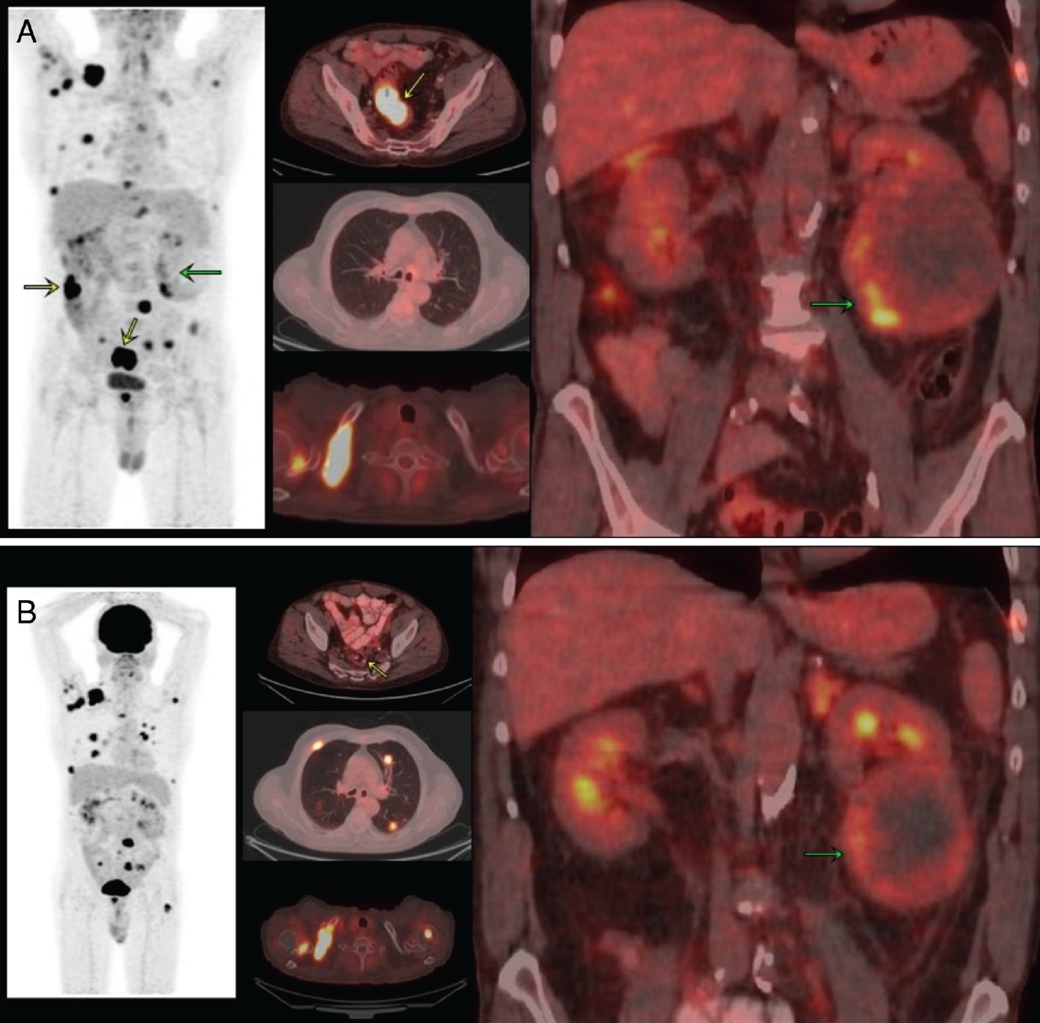
Figure 1
Figure 1. A) A 73-year-old man was referred for staging with 18F-fluorodeoxyglucose (18F-FDG) positron emission tomography/computed tomography (PET/CT) due to adenocarcinoma of the sigmoid colon. MIP and fusion images show two 18F-FDG-avid soft-tissue formations in the sigmoid and ascending colon [maximum standardized uptake value (SUVmax): 47.9]-yellow arrows. Also, a large and heterogeneous formation in the middle and lower third of the left kidney (87x95 mm), with increased 18F-FDG uptake in the soft tissue components of the lesion (SUVmax: 8.6) - green arrow. The scan showed multiple metabolically active lytic bone lesions with soft tissue components - ​​e.g., in the right clavicle, SUVmax: 15.8. 18F-FDG-negative small pulmonary nodules and mediastinal/hilar lymph nodes with mildly elevated metabolic activity were suspected as secondary foci. Considering these findings, the patient was referred for histological evaluation. The patient underwent surgery for the primary colonic lesions and partial resection of the tumor formation in the kidney. Given the low incidence of bone metastasis from colon carcinoma, even in the setting of a high initial T stage, as well as the macromorphological appearance of bone lesions, the latter was assumed to be associated with renal carcinoma. The registered lung nodules were left for observation due to their small size and lack of increased glucose metabolism on staging PET/CT. Multiple primary malignancies are increasingly seen in daily oncology practice, even though their frequency in the same or different organ systems is variable, ranging from 2% to 17% (1). Given the relatively low incidence of renal cell and colon carcinoma, reported at approximately 3.8% and 8.2% of total cancer cases diagnosed each year (2), the clinical scenario of co-occurring cancer is quite rare. According to the literature, in most cases, renal cell carcinoma (RCC) is associated with other primary malignancies, including prostate, bladder, and rectal cancers, as well as non-Hodgkin’s lymphoma (3). Regardless of the overall incidence of synchronous cancers, the most frequent occurrence is observed in the presence of initial colon/rectum in 2-5% of patients, followed by breast, lung, prostate, and urinary bladder tumors (4,5). Particularly in obstructive cancers, 18F-FDG PET/CT could be a valuable method for detecting synchronous colon tumors, with a high negative predictive value of 96.7% and accuracy of 87.5% (6). Diagnosis of second or more primary malignancies in patients with known cancer may be of significant importance for further therapeutic management (7) and can also accurately assess therapeutic response. In conclusion, PET/CT is a valuable hybrid technology that can more easily differentiate synchronous or metachronous tumors due to the combination of whole-picture visualization along with the different 18F-FDG uptake patterns in oncological diseases. B) Follow-up 18F-FDG PET/CT was performed after anterior rectal resection, transrectal polypectomy, and left kidney resection. Histological report was positive for 1. moderately differentiated adenocarcinoma of the rectum, pT3 pN0 LV0 Pn+ R0; 2. moderately differentiated adenocarcinoma of the cecum, pT1Nx, LV0 R0. 3. RCC, clear cell variant. The patient received targeted and osteomodulator therapy (sunitinib and denosumab). The MIP and axial and coronal fusion images of the follow-up scan show the metabolic and morphological progressive course of the disease, mainly involving lung and bone dissemination and persistence of metabolically active tumor formation with reduced size (due to partial resection).
View larger Download

Footnotes

Ethics

Informed Consent: Institutional review board approval was not required. Informed consent was obtained from each participant.

Authorship Contributions

Concept: P.N., V.H., Design: P.N., V.H., G.G., M.I., Data Collection or Processing: Y.G., S.V., G.G., E.R., Analysis or Interpretation: G.G., E.R., M.I., Literature Search: Y.G., S.V., G.G., Writing: P.N., V.H., M.I.

Conflict of Interest: No conflicts of interest were declared by the authors.

Financial Disclosure: The authors declare that this study has received no financial support.

References

1

Vogt A, Schmid S, Heinimann K, Frick H, Herrmann C, Cerny T, Omlin A. Multiple primary tumours: challenges and approaches, a review. ESMO Open. 2017;2:e000172.

2

Howlader N, Noone AM, Krapcho M, Garshell J, Miller D, Altekruse SF, Kosary CL, Yu M, Ruhl J, Tatalovich Z,Mariotto A, Lewis DR, Chen HS, Feuer EJ, Cronin KA (eds). SEER Cancer Statistics Review, 1975-2011, National Cancer Institute. Bethesda, MD, https://seer.cancer.gov/archive/csr/1975_2011/, based on November 2013 SEER data submission, posted to the SEER web site, April 2014. http://seer.cancer.gov/csr/1975_2011.

3

Rabbani F, Grimaldi G, Russo P. Multiple primary malignancies in renal cell carcinoma. J Urol. 1998;160:1255-1259.

4

Ringland CL, Arkenau HT, O’Connell DL, Ward RL. Second primary colorectal cancers (SPCRCs): experiences from a large Australian Cancer Registry. Ann Oncol. 2010;21:92-97.

5

Hayat MJ, Howlader N, Reichman ME, Edwards BK. Cancer statistics, trends, and multiple primary cancer analyses from the Surveillance, Epidemiology, and End Results (SEER) Program. Oncologist. 2007;12:20-37.

6

Maeda C, Endo S, Mori Y, Mukai S, Hidaka E, Ishida F, Kudo SE. The ability of positron emission tomography/computed tomography to detect synchronous colonic cancers in patients with obstructive colorectal cancer. Mol Clin Oncol. 2019;10:425-429.

7

Chun-Sing W, Nan-Jie G, Yiu-Ching C. Prevalence of synchronous second primary malignancy: identification using whole body PET/CT imaging. Clin Imaging. 2014;38:179-186.

