

Abstract
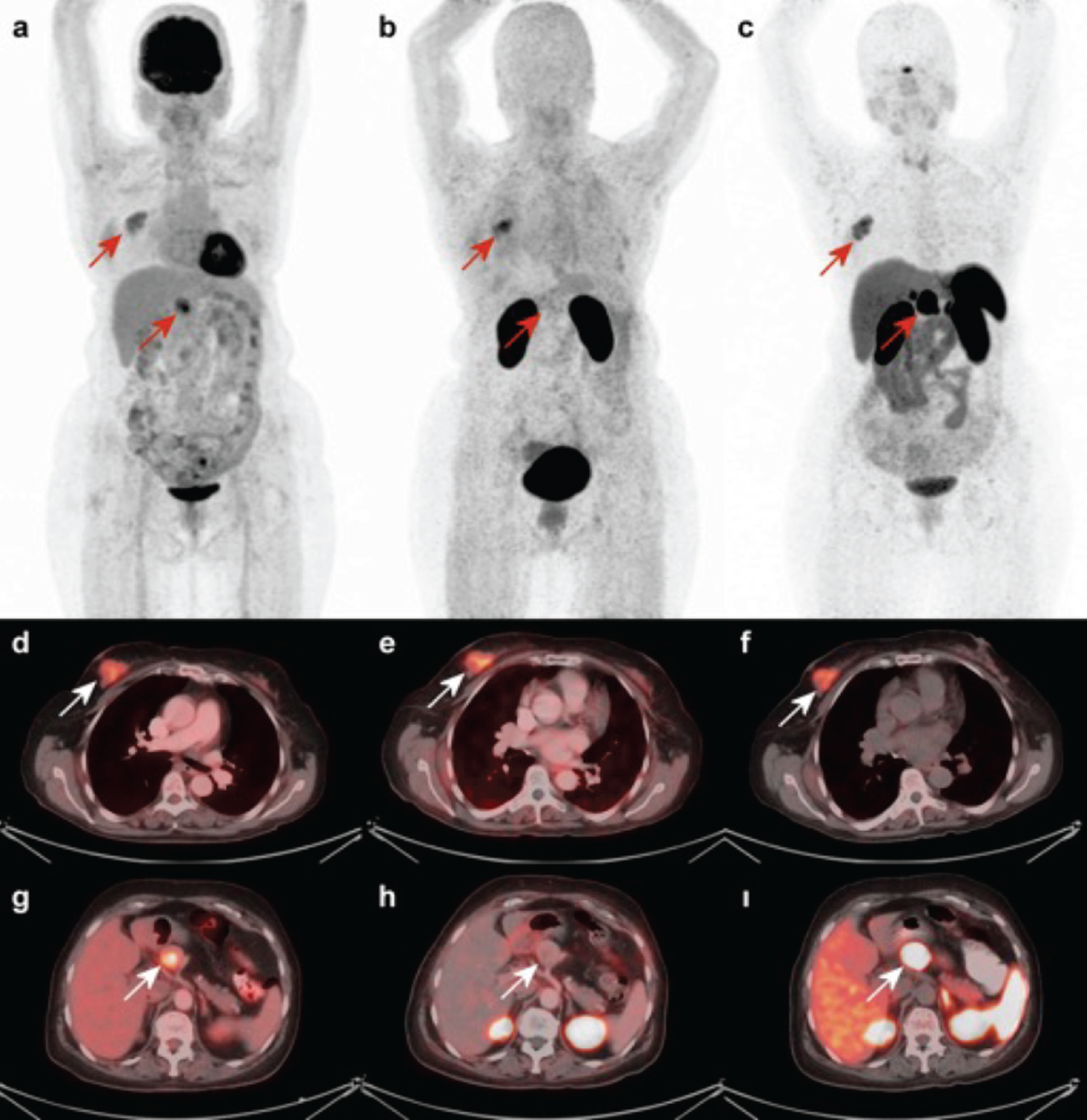
Integrins play crucial roles in the migration of tumor cells during angiogenesis and metastasis. Consequently, avβ6-integrin-targeted positron emission tomography (PET) radiopharmaceuticals have been developed and tested in humans, with clinical trials highlighting their applications in idiopathic pulmonary fibrosis and carcinomas. However, data on integrins are limited, and the role of [68Ga]Ga-Trivehexin tomography/computed tomography (CT) PET/CT is not well-established. Some studies have suggested that [68Ga]Ga-Trivehexin PET/CT is more specific than 18F-fluorodeoxyglucose (18F-FDG) PET/CT, which can yield false-positive results. It has been shown to be more efficient in evaluating pancreatic lesions and head and neck tumors. The role of [68Ga]Ga Trivehexin PET/CT in neuroendocrine tumors is not yet clearly defined. In our case, integrin was negative in the pancreatic neuroendocrine tumor but positive in the breast lobular tumor. Additionally, we observed that the lobular carcinoma lesion in the right breast is somatostatin receptor+positive on [68Ga]Ga-DOTATATE PET/CT.
Keywords: Neuroendocrine tumor, integrin positron emission tomography, [68Ga]Ga-Trivehexin positron emission tomography/computed tomography
Footnotes
Ethics
Informed Consent: Patient consent was obtained for this study.
Authorship Contributions
Surgical and Medical Practices: O.Y., M.K., Concept: G.B., N.A.S., K.A., L.K., Design: N.A.S., L.K., Data Collection or Processing: G.B., Analysis or Interpretation: G.B., N.A.S., L.K., Literature Search: N.A.S., K.A., Writing: G.B., N.A.S., K.A.
Conflict of Interest: No conflicts of interest were declared by the authors.
Financial Disclosure: The authors declare that they have received financial support for this study from Nukleon Nuclear Technology Research Ind. Trade Ltd. Co.
References
Urso L, Napolitano R, Speltri G, Tuncel M, Badrane I, Uccelli L, Porto F, Martini P, Niorettini A, Cittanti C, Bartolomei M, Boschi A. Ga-trivehexin: current status of avβ6-integrin imaging and perspectives. Cancers. 2025; 17:1504.
Maher TM, Simpson JK, Porter JC, Wilson FJ, Chan R, Eames R, Cui Y, Siederer S, Parry S, Kenny J, Slack RJ, Sahota J, Paul L, Saunders P, Molyneaux PL, Lukey PT, Rizzo G, Searle GE, Marshall RP, Saleem A, Kang’ombe AR, Fairman D, Fahy WA, Vahdati-Bolouri M. A positron emission tomography imaging study to confirm target engagement in the lungs of patients with idiopathic pulmonary fibrosis following a single dose of a novel inhaled avβ6 integrin inhibitor. Respir Res. 2020; 21:75.
Strobel O, Büchler MW. Pancreatic cancer: FDG-PET is not useful in early pancreatic cancer diagnosis. Nat Rev Gastroenterol Hepatol. 2013; 10:203-205.
Quigley NG, Steiger K, Hoberück S, Czech N, Zierke MA, Kossatz S, Pretze M, Richter F, Weichert W, Pox C, Kotzerke J, Notni J. PET/CT imaging of head-and-neck and pancreatic cancer in humans by targeting the “cancer integrin” avβ6 with Ga-68-Trivehexin. Eur J Nucl Med Mol Imaging. 2022; 49:1136-1147.
Alan Selçuk N, Akçay K, Beydağı G, Sönmez Ö, Çelik S, Öven BB, Kabasakal L. Revision of the histopathological examination following 68Ga-DOTA-FAPI-04 PET/CT of a breast tumor diagnosed as invasive ductal carcinomatosis. Mol Imaging Radionucl Ther. 2024; 33:134-137.

