

Abstract
Pancreatic exocrine carcinoma (PEC) is a lethal malignancy with high mortality rates because of its aggressive nature and frequent late-stage diagnosis. When histopathological diagnosis becomes unfeasible because of patient deterioration, clinicians must rely on clinical, biochemical, and radiological findings. This case report describes a 78-year-old woman with aggressive PEC visualized through 18F-fluorodeoxyglucose (18F-FDG) positron emission tomography/computed tomography. The imaging revealed an intensely hypermetabolic head mass, hepatic superscan, and hypermetabolic abdominal lymphadenopathy. Despite strong clinical indicators suggesting stage IV PEC, rapid disease progression and patient demise precluded histopathological confirmation, emphasizing the poor prognosis associated with hepatic superscan in this context.
Keywords: Pancreatic carcinoma, hepatic superscan, fulminant metastasis, 18F-FDG, PET/CT
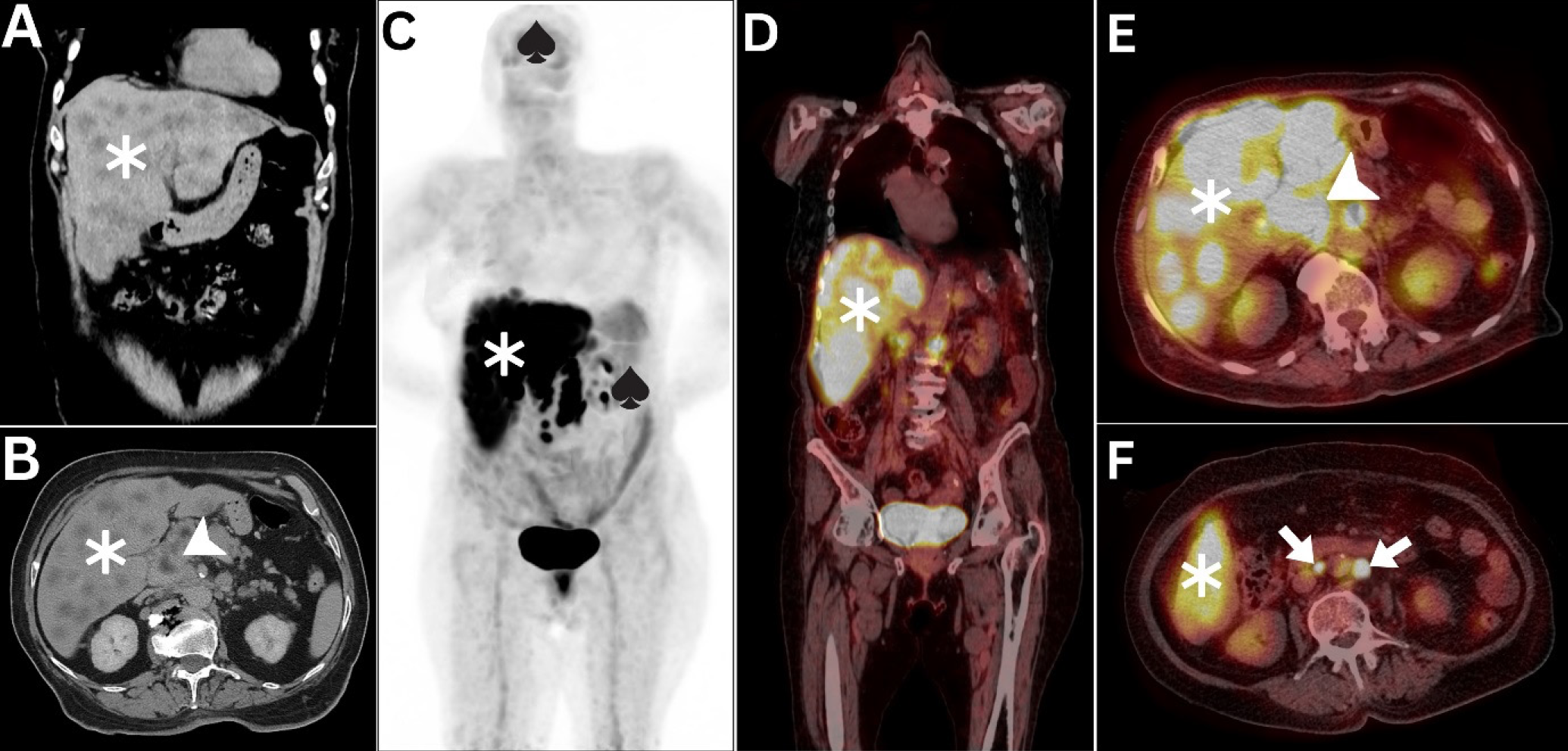
Hepatic superscan is a rare molecular imaging entity observed in aggressive hematologic and solid malignancies (,). To our knowledge, its occurrence in pancreatic carcinoma has not been previously reported. The distinct pattern of hepatic superscan characterized by intensely hypermetabolic hepatomegaly and suppressed 18F-FDG activity in organs with normally intensified 18F-FDG metabolism can be observed on MIP images (). Recent observations have highlighted the prognostic significance of hepatic superscan, associating it with poor prognosis and rapid onset of disease, often before management can be initiated. Such cases frequently receive delayed diagnosis because of neglect, delayed diagnosis, or misdiagnosis (,,,). Therefore, recognition of this unique neoplastic pattern should prompt immediate clinical and oncologic intervention to achieve the best possible outcome.
Footnotes
Ethic
Informed Consent: The patient consent was obtained.
Authorship Contributions
Surgical and Medical Practices: D.A., M.J., A.A., Concept: D.A., M.J., A.A., Design: D.A., M.J., A.A., Data Collection or Processing: D.A., M.J., A.A., Analysis or Interpretation: D.A., A.A., Literature Search: M.J., A.A., Writing: D.A., A.A.
Conflict of Interest: No conflicts of interest were declared by the authors.
Financial Disclosure: The authors declare that this study has received no financial support.
References
Basu S, Nair N. Unusually elevated liver radioactivity on F-18 FDG PET in Hodgkin’s disease: hepatic ‘superscan’. Clin Nucl Med. 2004;29:626-628.
Abdlkadir AS, Al-Adhami D, Alsyouf B, Alhouwari R, Al-Rasheed U, Jaber O, Mohamad I, Al-Ibraheem A. Hepatic Superscan in Medullary Thyroid Carcinoma: A Rare Presentation in [F]FDG PET/CT. Nucl Med Mol Imaging. 2024;58:147-149.
Abdlkadir AS, Almasri N, Estrada E, Fani Bozkurt M, Al-Ibraheem A. 18 F-FDG PET/CT Unveils Coexistent Myelomatous Hepatosplenic and Thyroid Cartilage Involvement : A Rare Presentation in Multiple Myeloma. Clin Nucl Med. 2024;49:897-898.
Taneja S, Achong DM. Hepatic superscan on PET/CT due to Richter’s transformation of chronic lymphocytic leukemia. Clin Nucl Med. 2010;35:644-645.
Yang G, Nie P, Wang Z, Xing X. (18)F-FDG hepatic superscan caused by a non-germinal center subtype of diffuse large B-cell lymphoma. Eur J Nucl Med Mol Imaging. 2016;43:1928.

