

Abstract
Bronchial carcinoids are low-grade neuroendocrine tumors with slow growth rates and the potential to spread to nearby lymph nodes. Here we present a challenging case of bronchial carcinoid visualized alongside an adjacent benign bronchocele. Chest computed tomography (CT) identified the endobronchial mass with unclear morphological and diagnostic insights. A differential diagnosis of several benign and malignant etiologies was made. Subsequently, an endobronchial biopsy confirmed lung carcinoid. For better evaluation, a 68Ga-labeled 1,4,7,10-tetraazacyclododecane-N,N’,N’’,N’’’-tetraacetic acid-d-Phe1-Tyr3-octreotide (68Ga-DOTATOC) positron emission tomography/CT scan was performed. The scan revealed a locally confined endobronchial mass with intense 68Ga-DOTATOC expression. Adjacent benign bronchocele was visualized with insignificant 68Ga-DOTATOC expression. Histopathological examination of the resected upper lobe confirmed these findings. This case highlights the importance of somatostatin receptor imaging in accurately identifying the extent of carcinoid tumors in the primary, nodal, and metastatic domains.
Keywords: Bronchocele, bronchial carcinoid, diagnostic challenge, 68Ga-DOTATOC, somatostatin receptor imaging, PET/CT
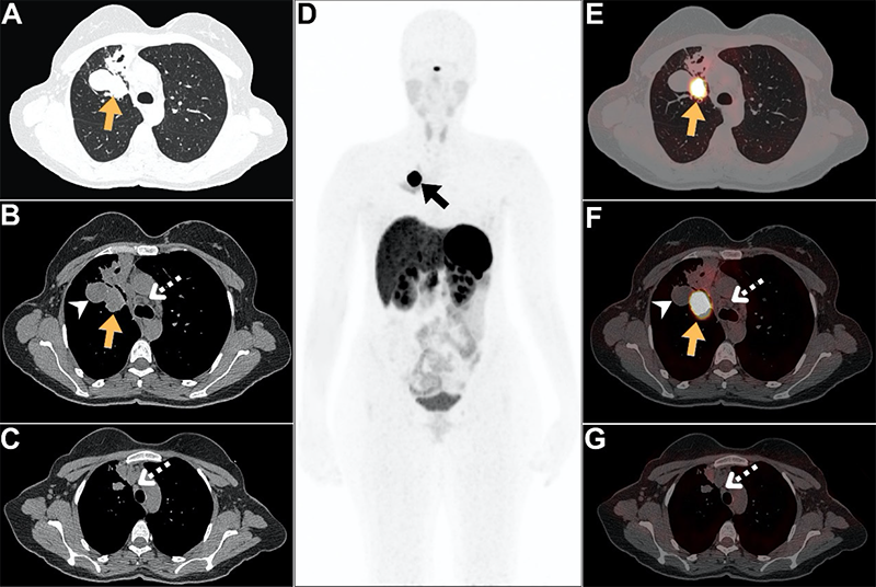
Bronchial carcinoids constitute 2% of all pulmonary malignancies (1). These tumors display various clinical, morphological, and biological patterns (1). Bronchial carcinoids may exhibit concurrent bronchocele in proximity (2). Bronchoceles, also termed bronchial mucocele, are typically benign but may masquerade as part of the primary tumor, leading to disease upstaging (2). CT evaluation alone may complicate precise tumor size determination due to density similarities and proximity (3). Occasionally, cystic malignancies were incorrectly identified by CT as mucocele or bronchocele (4,5). In such scenarios, SSTR imaging aids in accurate disease diagnosis. This unique case highlights the first instance in which 68Ga-DOTATOC PET/CT discriminated a bronchial carcinoid tumor from a subsequent bronchocele. These findings underscore the important role of SSTR imaging in distinguishing benign bronchocele from endobronchial carcinoid, ensuring precise staging across TNM.
Footnotes
Ethics
Informed Consent: Informed consent was obtained from the patient.
Authorship Contributions
Surgical and Medical Practices: A.A.-I., R.A., B.A., A.S.A., H.H., Concept: A.A.-I., R.A., B.A., A.S.A., Design: A.A.-I., R.A., B.A., Data Collection or Processing: A.A.-I., R.A., B.A., H.H., Analysis or Interpretation: A.A.-I., A.S.A., H.H., Literature Search: R.A., A.S.A., Writing: A.A.-I., A.S.A.
Conflict of Interest: No conflicts of interest were declared by the authors.
Financial Disclosure: The authors declare that this study has received no financial support.
References
Araujo-Castro M, Pascual-Corrales E, Molina-Cerrillo J, Moreno Mata N, Alonso-Gordoa T. Bronchial Carcinoids: From Molecular Background to Treatment Approach. Cancers (Basel). 2022;14:520.
Kakarla B. Bronchocele, a common but underrecognized condition: a systematic review. Monaldi Arch Chest Dis. 2022;93.
Yadav V, Rathi V. Bronchial carcinoid with bronchocele masquerading as Scimitar syndrome on chest radiograph. Radiol Case Rep. 2021;16:710-713.
Sandhu FA, Martuza RL. Craniofacial hemangiopericytoma associated with oncogenic osteomalacia: case report. J Neurooncol. 2000;46:241-247.
Shikhare SN. Imaging of Thoracic Malignancies. Thoracic Imaging. 2019:101-146.

