68Ga-PSMA-Avid Sinonasal Intestinal-Type Adenocarcinoma
PDF

Interesting Image

68Ga-PSMA-Avid Sinonasal Intestinal-Type Adenocarcinoma

Mol Imaging Radionucl Ther 2025;34(2):139-142 DOI: 10.4274/mirt.galenos.2024.48403
, , ,
PDF
Permissions

Abstract

A rare tumor, intestinal-type adenocarcinoma (ITAC), accounts for 8-25% of all sinonasal malignancies. The tumor’s histological resemblance to gastrointestinal tract adenocarcinoma is implied by its name. ITAC develops in the ethmoid sinus and upper nasal cavity. Herein, we present sinonasal ITAC with increased prostate-specific membrane antigen expression and fluorodeoxyglucose uptake.

Keywords: 68Ga-PSMA, sinonasal intestinal-type adenocarcinoma, fluorodeoxyglucose

,
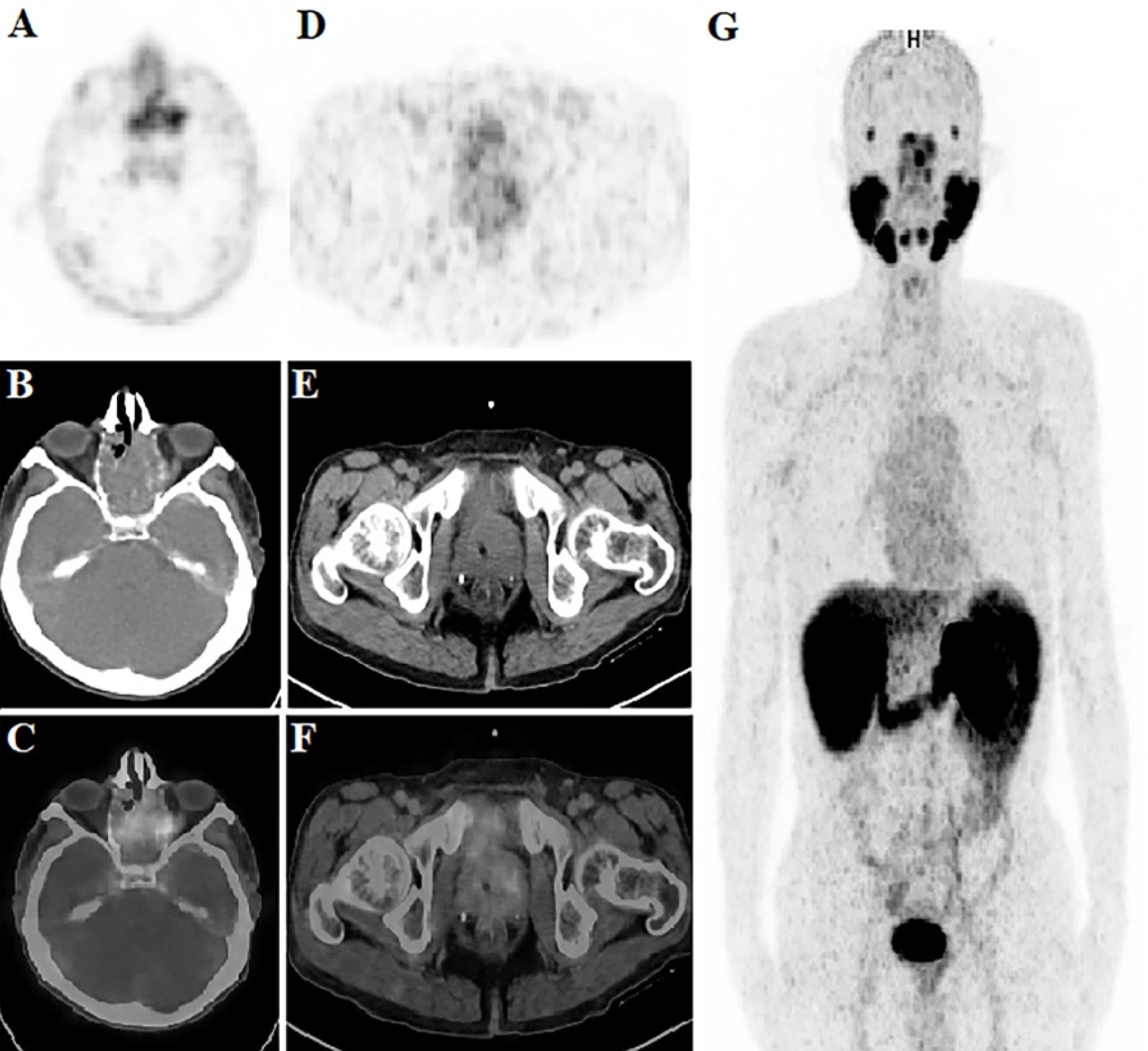
Figure 1
Figure 1. A systematic biopsy was conducted on the prostate gland of a 64-year-old man who previously underwent right radical nephrectomy for renal cell cancer due to the presence of high PSA (9.67 ng/mL). Biopsy revealed an International Society of Urological Pathology grade 3 prostate cancer with a Gleason score of 3+4. 68Ga-prostate-specific membrane antigen (68Ga-PSMA) positron emission tomography/computerized tomography (PET/CT) scan was conducted to assess the extent of metastatic disease. 68Ga-PSMA-avid sinonasal mass and heterogeneous 68Ga-PSMA uptake in the prostate gland were observed on the 68Ga-PSMA PET maximum intensity projection image (A). Axial 68Ga-PSMA PET (B), computed tomography (C), and fused 68Ga-PSMA PET/CT (D) images showing 68Ga-PSMA–avid mass involving the left maxillary sinus, left half of the nasal cavity, and both posterior ethmoid sinuses (SUVmax: 12.33). Axial 68Ga PSMA PET (E), CT (F), and fused 68Ga-PSMA PET/CT (G) images showed heterogeneous slightly increased uptake of 68Ga-PSMA in the large prostate glands (SUVmax: 7.33). The histomorphology of the biopsy from the sinonasal mass revealed stratified papillary structures consisting of atypical cells. It was observed that these cells were immunohistochemically stained with CK7, CK20, SATB-2, and Ki-67 (high), but not with PSA and NKX3.1, and these findings were evaluated as compatible with sinonasal intestinal-type adenocarcinoma (ITAC).
View larger Download
Figure 2
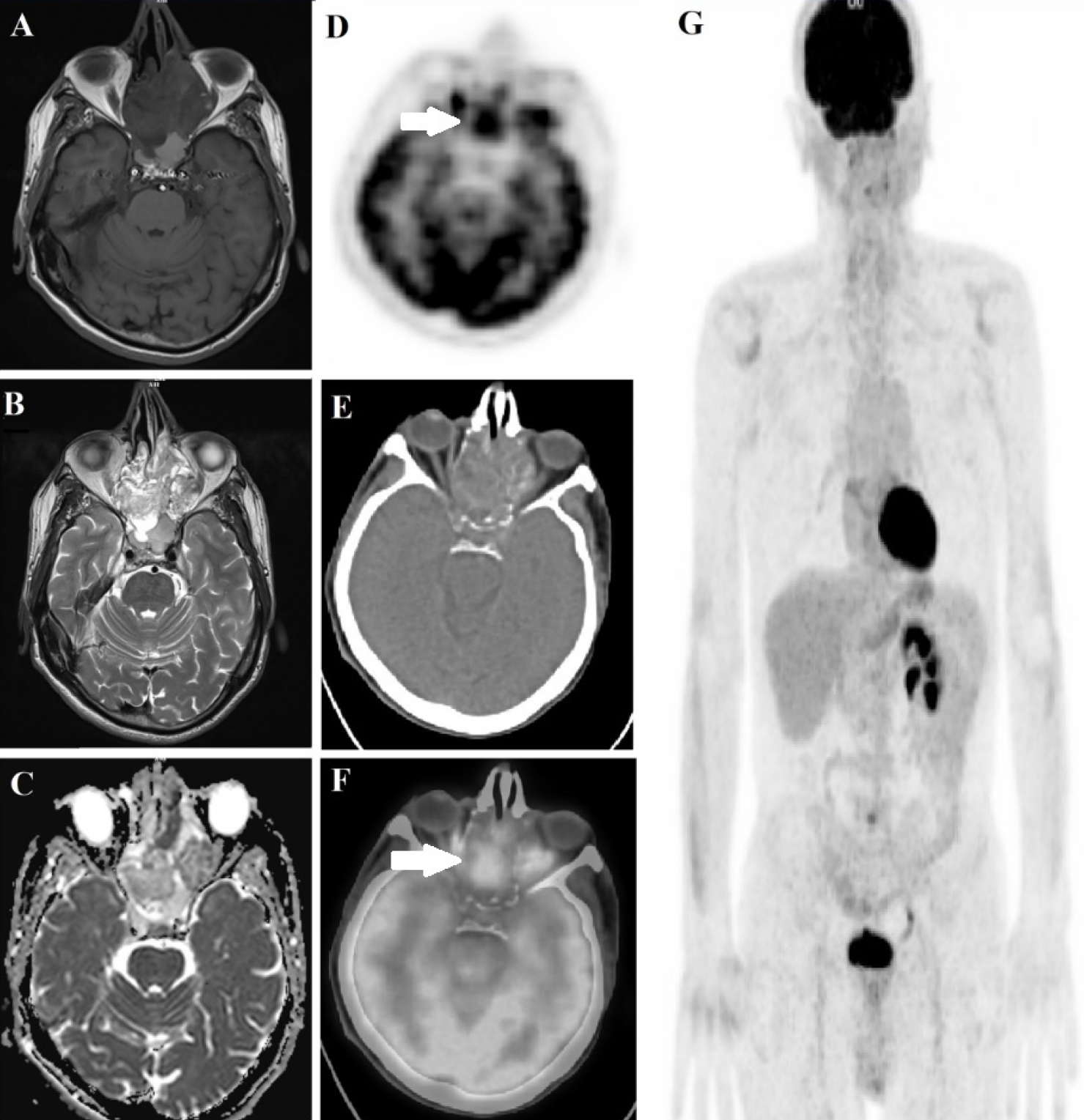
Figure 2. To rule out the risk of metastasis from other adenocarcinomas, 18F- fluorodeoxyglucose (FDG) PET/CT was performed. Only the sinonasal mass was found to have 18F- FDG avidity. Axial 18F-FDG) PET (E), CT (F), and fused 18F-FDG) PET/CT (G) images showing marked hypermetabolism (SUVmax: 12.18) in the primary tumor. There was no lymph node involvement in the cervical region, evidence of another primary adenocarcinoma, or distant metastasis. In addition, the right kidney was not observed after the operation. Magnetic resonance imaging shows lesions with low signal on T1-weighted images (A), high signal on T2-weighted images (B), and diffusion-weighted images (C). The upper nasal cavity and ethmoid sinus are the primary origin sites of ITACs (,,). The most common symptoms include unilateral, non-spesific unilateral nasal blockage, epistaxis, and rhinorrhea (). Colonic ITAC is the most common subtype, with papillary, mucinous, solid, and mixed types (). It is difficult to distinguish between primary ITAC and nasal cavity metastases of adenocarcinomas due to the similarity of microscopic characteristics and immunohistochemistry (,). Therefore, diagnosing primary ITAC in the sinonasal region is only possible after excluding all other adenocarcinomas. This case hints at the value of 18F-FDG PET/CT in the diagnosis of primary ITAC in the nasal cavity and demonstrates the 68Ga-PSMA avidity of ITAC. In general, for non-prostatic solid tumors associated with neovascularity, endothelial PSMA expression has been shown (). Therefore, PSMA expression in non-prostatic solid tumors on PET imaging requires readers to consider the histology, imaging, and clinical details of tumor neovascularization. Although demonstrating PSMA expression in non-prostatic diseases is disadvantageous, PSMA has paved the way for PET imaging to be applied as an additional theranostic tool for this malignancy ().
View larger Download

Footnotes

Informed Consent: Patient consent was obtained.

Authorship Contributions

Surgical and Medical Practices: H.Ö., H.Öz., M.F.G., O.Ö.E., Concept: H.Ö., H.Öz., M.F.G., O.Ö.E., Design: H.Ö., H.Öz., M.F.G., O.Ö.E., Data Collection or Processing: H.Ö., H.Öz., M.F.G., O.Ö.E., Analysis or Interpretation: H.Ö., H.Öz., M.F.G., O.Ö.E., Literature Search: H.Ö., H.Öz., M.F.G., O.Ö.E., Writing: H.Ö., H.Öz., M.F.G., O.Ö.E.

Conflict of Interest: No conflicts of interest were declared by the authors.

Financial Disclosure: The authors declare that this study has received no financial support.

References

1

Kılıç S, Samarrai R, Kılıç SS, Mikhael M, Baredes S, Eloy JA. Incidence and survival of sinonasal adenocarcinoma by site and histologic subtype. Acta Otolaryngol. 2018;138:415-421.

2

Abecasis J, Viana G, Pissarra C, Pereira T, Fonseca I, Soares J. Adenocarcinomas of the nasal cavity and paranasal sinuses: a clinicopathological and immunohistochemical study of 14 cases. Histopathology. 2004;45:254-259.

3

Hoeben A, van de Winkel L, Hoebers F, Kross K, Driessen C, Slootweg P, Tjan-Heijnen VC, van Herpen C. Intestinal-type sinonasal adenocarcinomas: The road to molecular diagnosis and personalized treatment. Head Neck. 2016;38:1564-1570.

4

Choussy O, Ferron C, Védrine PO, Toussaint B, Liétin B, Marandas P, Babin E, De Raucourt D, Reyt E, Cosmidis A, Makeiff M, Dehesdin D; GETTEC Study Group. Adenocarcinoma of Ethmoid: a GETTEC retrospective multicenter study of 418 cases. Laryngoscope. 2008;118:437-443.

5

Vivanco B, Llorente JL, Perez-Escuredo J, Alvarez Marcos C, Fresno MF, Hermsen MA. Benign lesions in mucosa adjacent to intestinal-type sinonasal adenocarcinoma. Patholog Res Int. Patholog Res Int. 2011;2011:230147.

6

Projetti F, Durand K, Chaunavel A, Léobon S, Lacorre S, Caire F, Bessède JP, Moreau JJ, Coulibaly B, Labrousse F. Epidermal growth factor receptor expression and KRAS and BRAF mutations: study of 39 sinonasal intestinal-type adenocarcinomas. Hum Pathol. 2013;44:2116-2125.

7

Fu Z, Zhang J, Liu M, Li Z, Li Q. Diagnosis of Primary Intestinal-Type Adenocarcinoma in the Nasal Cavity by 18F-FDG PET/CT. Clin Nucl Med. 2016;41:888-889.

8

Backhaus P, Noto B, Avramovic N, Grubert LS, Huss S, Bögemann M, Stegger L, Weckesser M, Schäfers M, Rahbar K. Targeting PSMA by radioligands in non-prostate disease-current status and future perspectives. Eur J Nucl Med Mol Imaging. 2018;45:860-877.

9

An S, Huang G, Liu J, Wei W. PSMA-targeted theranostics of solid tumors: applications beyond prostate cancers. Eur J Nucl Med Mol Imaging. 2022;49:3973-3976.