

Abstract
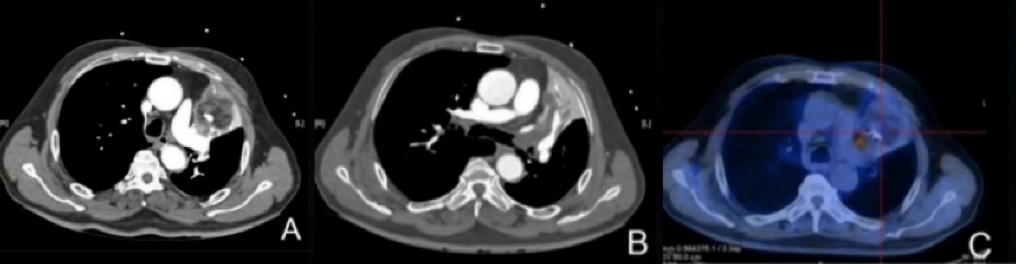
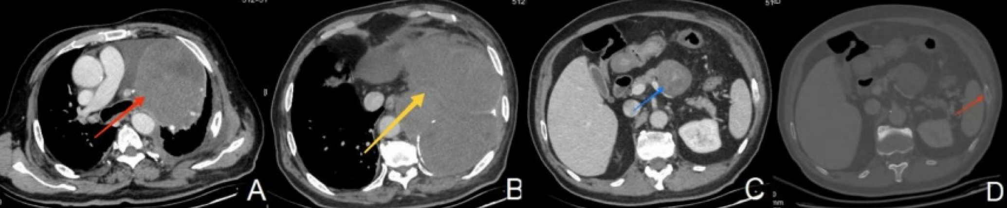
Primary liposarcoma of the lung is extremely rare. To date, only 24 cases have been reported in the English literature. Herein, we present a case of well-differentiated pulmonary liposarcoma that was misdiagnosed as teratoma on positron emission tomography/computed tomography (CT) and contrast-enhanced CT. Radical surgery with left superior lobectomy and mediastinal lymph node dissection were performed. The patient experienced recurrence and distant metastases 33 months after surgery. He was alive at the time of writing this report (36 months postoperatively). To our knowledge, this is the first case report of pulmonary well-differentiated liposarcoma.
Keywords: Primary pulmonary liposarcoma, positron emission tomography/computed tomography, well-differentiated liposarcoma
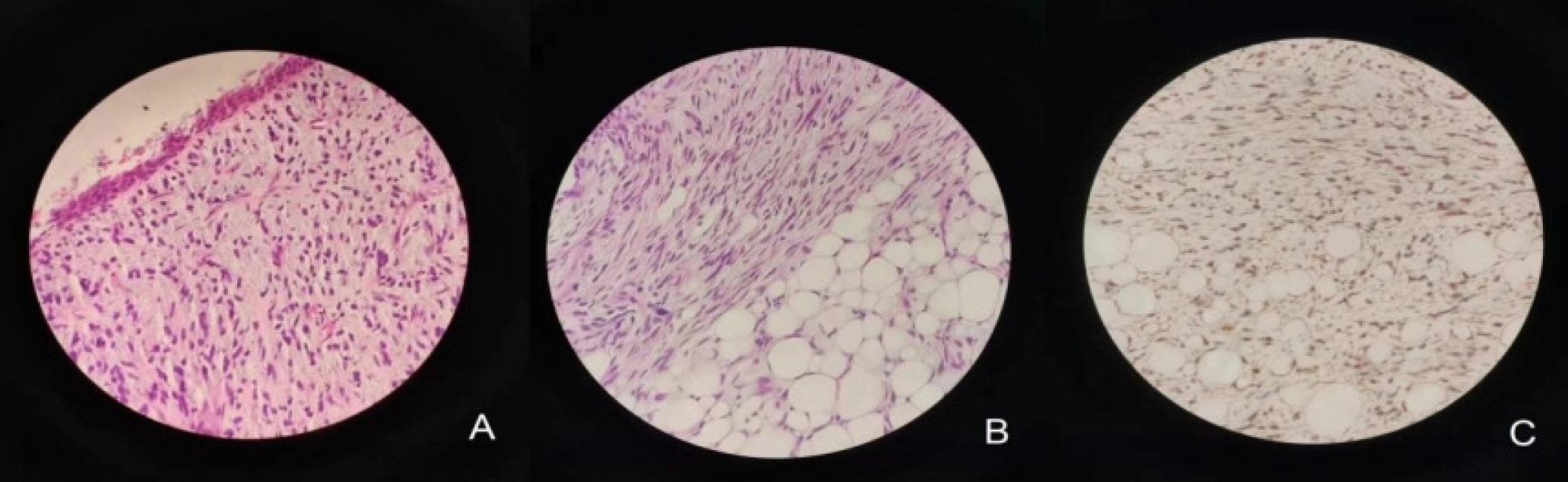
Primary liposarcoma of the lung is an extremely rare malignancy, accounting for approximately 0.2% of all pulmonary tumors (). Lipomatosis and asbestosis may be pathogenic factors (); According to the World Health Organization Classification of Soft Tissue Tumors (2020 edition), liposarcomas can be divided into five types: well-differentiated, dedifferentiated, myxoid, pleomorphic, and mixed (); Well-differentiated liposarcoma tumors contain a large amount of fat, usually ≥ 75% of the tumor volume (). Although well-differentiated liposarcomas have certain imaging characteristics, they still need to be differentiated from other lung tumors, especially those with fatty components such as teratomas, hamartomas, and other mesenchymal-derived tumors (). Preoperative imaging examination is difficult to correctly diagnose, and the final diagnosis depends on the immunohistochemical results. Immunohistochemistry of well-differentiated liposarcoma was positive for Vimentin, S-100, MDM2, and CDK4 (). The preferred treatment for primary pulmonary liposarcoma is radical resection and lymph node dissection (, ). Well-differentiated liposarcoma was the least invasive of the five pathological subtypes. The 5-year survival rate of well-differentiated liposarcomas is 87.1% () and the recurrence rate is 40-50% (). The median survival time of patients with primary intrathoracic liposarcoma according to the well-differentiated subtypes was 174 months ().
Footnotes
Ethics
Informed Consent: An informed consent was obtained from the patient.
Authorship Contributions
Concept: H.L., Design: H.L., Data Collection or Processing: Z.L., Analysis or Interpretation: Z.L., Literature Search: Z.L., Writing: H.L.
Conflict of Interest: No conflicts of interest were declared by the authors.
Financial Disclosure: The authors declare that this study has received no financial support.
References
Li B, Xin Z, Li Z, Zhang X. Giant pulmonary pleomorphic liposarcoma: A case report and literature review. Asian J Surg. 2023;46:1109-1110.
Dey T, Khosla D, Kumar D, Chatterjee D, Madan R, Singh H, Singh H, Kapoor R. Rare case of primary pulmonary pleomorphic liposarcoma treated with multimodal therapy. Ochsner J. 2021;21:431-435.
Choi JH, Ro JY. The 2020 WHO classification of tumors of soft tissue: selected changes and new entities. Adv Anat Pathol. 2021;28:44-58.
Said M, Migaw H, Hafsa C, Braham R, Golli M, Moussa A, Zakhama A, Elkamel A, Gannouni A. Imaging features of primary pulmonary liposarcoma. Australas Radiol. 2003;47:313-317.
Stark P, Eber CD, Jacobson F. Primary intrathoracic malignant mesenchymal tumors: pictorial essay. J Thorac Imaging. 1994;9:148-155.
Oh YJ, Yi SY, Kim KH, Cho YJ, Beum SH, Lee YH, Suh JS, Hur H, Kim KS, Kim SH, Choi YD, Shin KH, Jun HJ, Kim SJ, Lee J, Park SH, Noh SH, Rha SY, Kim HS. Prognostic model to predict survival outcome for curatively resected liposarcoma: a multi-institutional experience. J Cancer. 2016;7:1174-80.
Henricks WH, Chu YC, Goldblum JR, Weiss SW. Dedifferentiated liposarcoma: a clinicopathological analysis of 155 cases with a proposal for an expanded definition of dedifferentiation. Am J Surg Pathol. 1997;21:271-281.
Fu Z, Yang K, Yang X, Chen S, Wang W, Chen D, Zhao J, Li Z, Feng Q, Zhou Z, Wang L, Gao S, Liang J. Primary intrathoracic liposarcoma: a clinical analysis of 31 cases. Cancer Commun (Lond). 2019;39:15.

