Nasolacrimal Metastasis from Parotid Ductal Carcinoma Detected by 18F-FDG PET/CT
PDF

Interesting Image

Nasolacrimal Metastasis from Parotid Ductal Carcinoma Detected by 18F-FDG PET/CT

Mol Imaging Radionucl Ther 2025;34(1):82-84 DOI: 10.4274/mirt.galenos.2024.46667
, , , ,
PDF
Permissions

Abstract

A 39-year-old woman presented with left neck masses for 4 months and epiphora of the left eye for 3 weeks. Ultrasonography revealed a mass in the left parotid gland and multiple cervical lymph nodes. Biopsy of the mass in the left parotid gland revealed infiltrating ductal carcinoma. 18F-fluorodeoxyglucose positron emission tomography/computed tomography scan was undertaken, which showed a mass in the left parotid gland and multiple cervical lymph nodes with high metabolism. A nodule in the left nasolacrimal duct with high metabolism was observed. The nodule was surgically removed and pathologically confirmed as metastatic parotid ductal carcinoma.

Keywords: Nasolacrimal tumor, metastasis, parotid ductal carcinoma, 18F-FDG, PET/CT
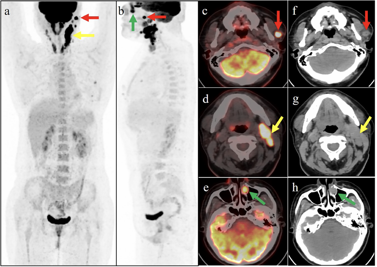
Figure 1
Figure 1. A 39-year-old woman presented with left neck masses for 4 months and epiphora of the left eye for 3 weeks. Ultrasonography revealed a mass in the left parotid gland and multiple cervical lymph nodes. The biopsy pathology of the mass in the left parotid gland revealed infiltrating ductal carcinoma. 18F-florodeoksiglukoz (18F-FDG) positron emission tomography/computed tomography scan was then performed for staging. The maximum-intensity projection image (a: anteroposterior) shows some areas of intense 18F-FDG activity in the left parotid region with an SUVmax of 24.0 (red arrow) and the left side of neck with SUVmax of 22.3 (yellow arrow). The left lateral MIP image (b) shows a focus of intense activity behind the nose, with an SUVmax of 17.3 (green arrow). On the axial images (c-e: fused PET/CT; f-h: CT), the red arrow corresponded well to the mass of the left parotid, the yellow arrow pointed to multiple cervical lymph nodes, and the green arrow corresponded to the nodule of the left nasolacrimal duct, which was surgically removed. Subsequently, the lesion was pathologically and immunohistochemically confirmed as metastatic parotid ductal carcinoma.
View larger Download

Figure 2
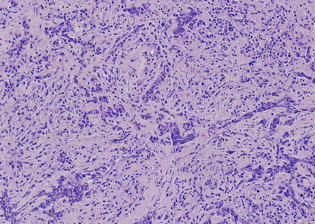
Figure 2. Histologically, the nasolacrimal duct specimen (hematoxylin and eosin stain, original magnification x200) showing mesenchyma infiltrated by strip-shaped invasive ductal carcinoma cells with the typical feature of irregular nested infiltrate. The most common clinical symptoms of lacrimal sac and duct tumors are epiphora, recurrent dacryocystitis, epistaxis, and/or lacrimal sac mass. These non-specific clinical manifestations often lead to the misdiagnosis of lacrimal sac tumors such as dacryocystitis (1). Tumors of the lacrimal sac and duct are divided into primary and secondary tumors. Primary malignant epithelial neoplasms include squamous cell carcinoma (SCC), transitional cell carcinoma, adenocarcinoma, adenoid cystic carcinoma, poorly differentiated carcinoma, and primary melanomas (2,3). The most common malignant lacrimal sac tumors are of epithelial origin, with the majority being SCC (4,5,6). Secondary tumors can occur from any cutaneous or paranasal sinus lesion or from distant organs, in the case of metastatic tumors, and may include carcinomas or melanomas (7,8). Our case indicates that metastatic tumors should be considered when high 18F-FDG accumulation is observed in the nodule of the lacrimal duct, especially when accompanied by other abnormal 18F-FDG uptake lesions.
View larger Download

Footnotes

Ethics

Informed Consent: Written informed consent has been obtained from the patient.

Authorship Contributions

Surgical and Medical Practices: J.L., Concept: Y.Z., Design: Y.Z., Data Collection or Processing: G.F., Analysis or Interpretation: Y.Z., Literature Search: X.G., Writing: K.F.

Conflict of Interest: No conflict of interest was declared by the authors.

Financial Disclosure: There were no sources of funding for this work.

References

1

Lee LN, Scott AR, Chan AW, Frankenthaler RA. Management of transitional cell carcinoma of the lacrimal sac: a multidisciplinary approach to orbit sparing treatment. Laryngoscope. 2010;120(Suppl 4):161.

2

El-Sawy T, Frank SJ, Hanna E, Sniegowski M, Lai SY, Nasser QJ, Myers J, Esmaeli B. Multidisciplinary management of lacrimal sac/nasolacrimal duct carcinomas. Ophthalmic Plast Reconstr Surg. 2013;29:454-457.

3

Subramaniam SS, Anand R, Mellor TK, Brennan PA. Primary Lacrimal Sac Melanoma With Metastatic Cervical Disease: A Review of the Literature and Case Report. J Oral Maxillofac Surg. 2017;75:1438-1441.

4

Zhang R, Qian J, Meng F, Yuan Y, Bai M, Bi Y, Xu B, Yuan Y, Hong R. A Staged Procedure in the Treatment of Primary Lacrimal Sac Epithelial Malignancy: A Retrospective Cases Analysis. Ophthalmic Plast Reconstr Surg. 2019;35:187-192.

5

Song X, Wang J, Wang S, Wang W, Wang S, Zhu W. Clinical analysis of 90 cases of malignant lacrimal sac tumor. Graefes Arch Clin Exp Ophthalmol. 2018;256:1333-1338.

6

Kumar VA, Esmaeli B, Ahmed S, Gogia B, Debnam JM, Ginsberg LE. Imaging Features of Malignant Lacrimal Sac and Nasolacrimal Duct Tumors. AJNR Am J Neuroradiol. 2016;37:2134-2137.

7

Sia KJ, Tang IP, Kong CK, Tan TY. Nasolacrimal relapse of nasopharyngeal carcinoma. J Laryngol Otol. 2012;126:847-850.

8

Ding J, Sun H, Xin Y, Li D. Metastatic hepatocellular carcinoma mimicking acute dacryocystitis. Can J Ophthalmol. 2018;53:132-134.