

Abstract
Neoplastic infiltration of the omentum is mostly caused by metastatic ovarian, gastric, colon, or pancreatic cancer. Lymphomatous infiltration of the omentum is rare because the omentum lacks a lymphoid component. To date, lymphomatous involvement of the omentum has only been reported in patients with non-Hodgkin lymphoma. Peritoneal lymphomatosis remains a rare presentation of malignant lymphoma characterized by diffuse peritoneal lesions and is frequently accompanied by ascites and mesenteric lesions. In this review, we aimed to illustrate the case of a 72 year old mal patient diagnosed with aggressive large B-cell lymphoma, adressed for initial extension assessment in whom 18F-fluorodeoxyglucose positron emission tomograph/computed tomography found unusual omental and mesenteric involvement.
Keywords: 18F-FDG PET-CT, non-Hodgkin’s lymphoma, omental cake
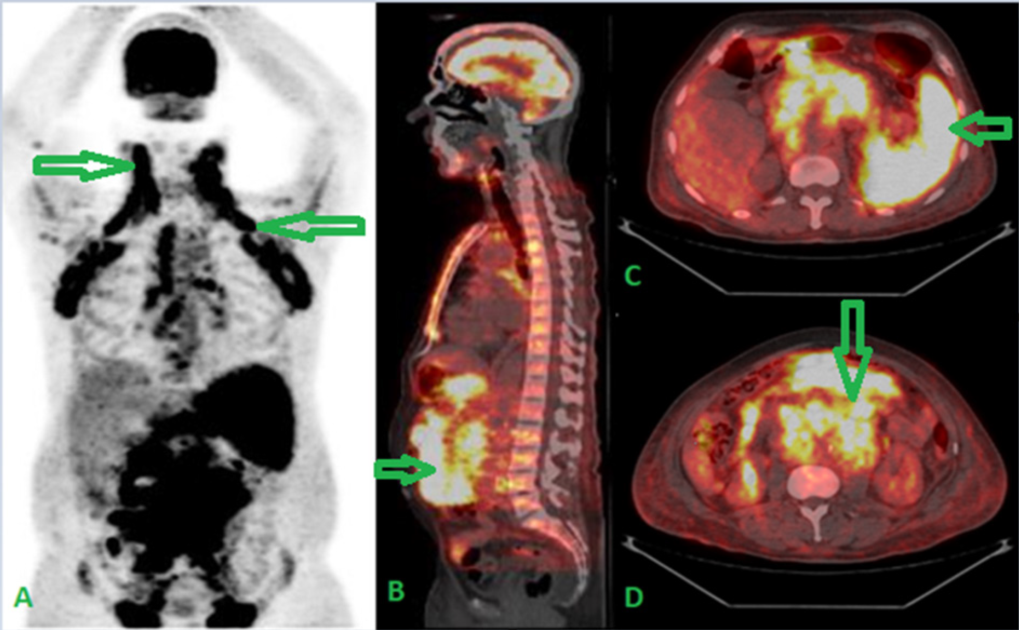
The greater omentum is a fibrous, fatty membrane structure formed by the continuation of the peritoneal surface of the anterior and posterior viscera. It extends downward from the stomach and folds in on itself, covering the small intestine and attaching to the upper side of the transverse colon (1).
Neoplastic infiltration of the omentum is most commonly caused by metastatic ovarian, gastric, colon, or pancreatic cancer. Lymphomatous infiltration of the omentum is rare because the omentum lacks a lymphoid component. To date, lymphomatous involvement of the omentum has only been reported in patients with non-Hodgkin lymphoma (1). Since most lymphomas are usually metabolically active, PET using 18F-FDG is very useful for early diagnosis and staging (2). PET-CT may be useful in the assessment of lymphomatous peritoneal tumor infiltration, as outlined in several published studies of other malignancies (3). Peritoneal lymphoma is radiologically characterized by diffuse peritoneal thickening and mass, and is associated with ascites in most cases. Intestinal wall thickening, retroperitoneal lymphadenopathy, and hepatosplenomegaly are also frequently observed (4). Peritoneal involvement is critical for accurate staging and treatment planning, as peritoneal involvement can be cured with appropriate therapeutic intervention and may result in death if treatment is delayed (5), which in our case highlights the value and the performances of PET-CT in initial staging and evaluation of the extent of the disease.
Footnotes
Ethics
Informed Consent: An informed consent was obtained from the patient.
Footnote
Concept: M.O., S.O.N., O.A.S., Y.B., A.D., Design: M.O., S.O.N., O.A.S., Y.B., A.D., Data Collection or Processing: M.O., Analysis or Interpretation: M.O., S.O.N., A.D., Literature Search: M.O., O.A.S., Writing: M.O.
Conflict of Interest: No conflicts of interest were declared by the authors.
Financial Disclosure: The authors declare that this study has received no financial support.
References
Jacobs JE, Salhany KE, Fox KR, Birnbaum BA. Omental caking in Hodgkin’s disease. Computed tomography findings. Clin Imaging. 1996;20:253-255.
Hutchings M, Barrington SF. PET/CT for therapy response assessment in lymphoma. J Nucl Med. 2009;50:21S-30S.
Turlakow A, Yeung HW, Salmon AS, Macapinlac HA, Larson SM. Peritoneal carcinomatosis: role of (18)F-FDG PET. J Nucl Med. 2003;44:1407-1412.
Karaosmanoglu D, Karcaaltincaba M, Oguz B, Akata D, Ozmen M, Akhan O. CT findings of lymphoma with peritoneal, omental and mesenteric involvement: peritoneal lymphomatosis. Eur J Radiol. 2009 2009;71:313-317.
Ichikawa S, Fukuhara N, Saito K, Onodera K, Onishi Y, Yokoyama H, Ichinohasama R, Harigae H. Diffuse Large B-cell Lymphoma Presenting as Peritoneal Lymphomatosis: A Case Report and Literature Review. Intern Med. 2022;61:2057-2060.

