

Abstract
A 73-year-old woman with known diagnosis of chronic hepatitis B infection referred for 18F-fluorodeoxyglucose positron emission tomography/ computed tomography (18F-FDG PET/CT) and magnetic resonance imaging (MRI) after detection of a liver mass with 9 cm diameter in contrast enhanced CT. However, 18F-FDG PET/CT and MRI revealed diffuse infiltrating hepatocellular carcinoma lesions other than previously defined mass. After 9 months of Sorafenib treatment serum alpha feto protein levels dropped from 60,500 ng/mL to 801 ng/mL. Later 18F-FDG PET/MRI was performed for evaluation of response to treatment and revealed marked response to treatment.
Keywords: Hepatocellular carcinoma, positron emission tomography, fluorodeoxyglucose, magnetic resonance imaging, sorafenib
HCC is one of the most prevalent cancers in the world (,). In advanced HCC cases treatment choices can be limited to systemic treatments such as tyrosine-kinase receptor inhibitors, vascular endothelial growth factor inhibitors and immunotherapy agents (,,). As one of the most commonly used tyrosine-kinase inhibitors, sorafenib can increase overall survival in HCC patients (). It has been shown that pretreatment 18F-FDG PET/CT parameters have a prognostic value in HCC patients treated with sorafenib but intensity of 18F-FDG uptake may not predict response to sorafenib treatment (,). In this case the lesions previously undetected in CT were demonstrated with both components of PET/MRI. Additionally, 18F-FDG PET revealed different characteristics of these two pathologies, one being solitary non-FDG avid mass, other being intensely FDG-avid infiltrating lesions. Furthermore, more 18F-FDG avid infiltrating lesions had a marked response to sorafenib while the other large solid mass was merely exhibiting stable disease. An effective response to sorafenib is rare, and in this case both PET and MRI revealed dramatic response of infiltrating lesions to treatment, consistent with clinical findings. In conclusion, PET can reveal metabolic characteristics of HCC lesions and usage of high soft tissue contrast of MRI can help identify corresponding lesions in liver. This could make the usage of PET/MRI valuable for diagnosis, staging and as demonstrated above evaluation of response to treatment.
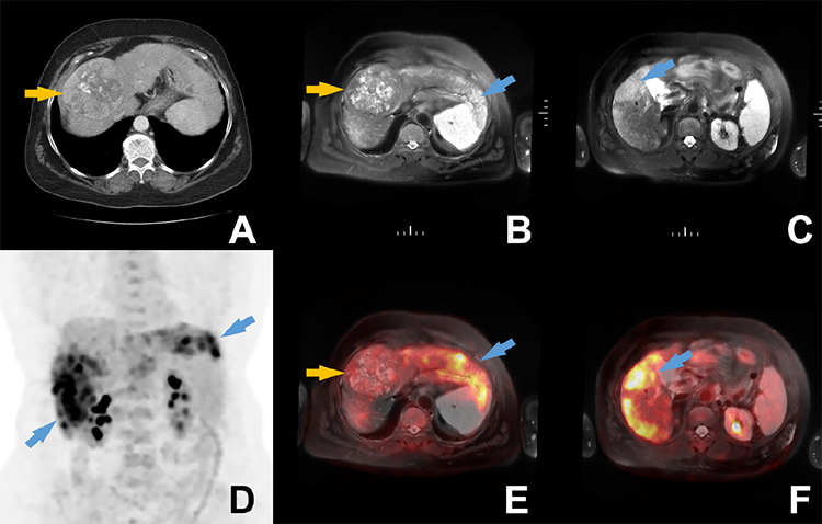
PET/CT MRI fusion images (E, F). Additionally, the previously defined 9 cm lesion (orange arrows) had very mild 18F-FDG uptake. Sorafenib treatment was initiated after the detection of advanced disease.
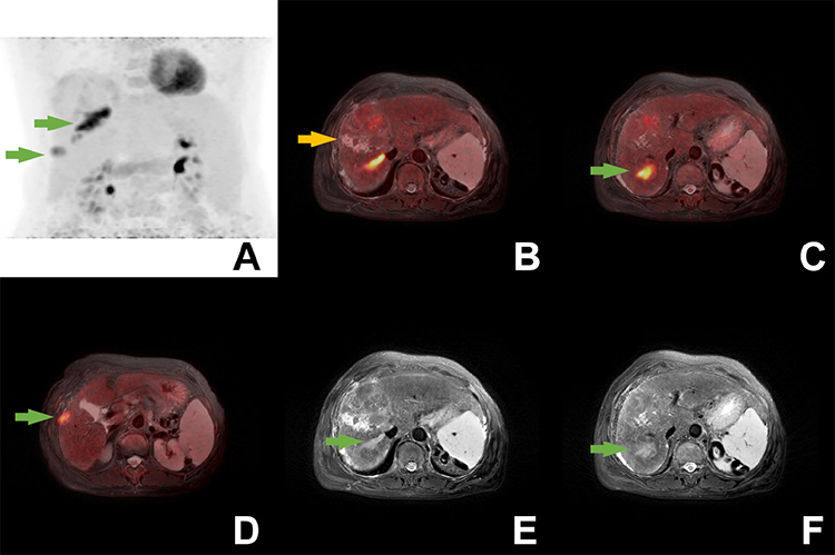
Footnotes
Ethics
Informed Consent: Written informed consent was obtained from the patient.
Authorship Contributions
Surgical and Medical Practices: E.Ö., B.D., D.K.Ö., Y.Ü., N.Ö.K., Concept: E.Ö., B.D., Data Collection or Processing: E.Ö., B.D., D.K.Ö., Y.Ü., N.Ö.K., Analysis or Interpretation: E.Ö., B.D., D.K.Ö., N.Ö.K., Literature Search: E.Ö., B.D., Writing: E.Ö., B.D.
Conflict of Interest: No conflicts of interest were declared by the authors.
Financial Disclosure: The authors declare that this study has received no financial support.
References
Sung H, Ferlay J, Siegel RL, Laversanne M, Soerjomataram I, Jemal A, Bray F. Global Cancer Statistics 2020: GLOBOCAN Estimates of Incidence and Mortality Worldwide for 36 Cancers in 185 Countries. CA Cancer J. Clin 2021;71:209-249.
El-Serag HB, Mason AC. Rising incidence of hepatocellular carcinoma in the United States. N Engl J Med. 1999;340:745-750.
Reig M, Forner A, Rimola J, Ferrer-Fàbrega J, Burrel M, Garcia-Criado Á, Kelley RK, Galle PR, Mazzaferro V, Salem R, Sangro B, Singal AG, Vogel A, Fuster J, Ayuso C, Bruix J. BCLC strategy for prognosis prediction and treatment recommendation: The 2022 update. J Hepatol. 2022;76:681-693.
European Association For The Study Of The Liver; European Organisation For Research And Treatment Of Cancer. EASL-EORTC clinical practice guidelines: management of hepatocellular carcinoma. J Hepatol. 2012;56:908-943.
Forner A, Reig M, Bruix J. Hepatocellular carcinoma. Lancet. 2018;391:1301-1314.
Fan G, Wei X, Xu X. Is the era of sorafenib over? A review of the literature. Ther Adv Med Oncol. 2020;12:1758835920927602.
Sung PS, Park HL, Yang K, Hwang S, Song MJ, Jang JW, Choi JY, Yoon SK, Yoo IR, Bae SH. 18F-fluorodeoxyglucose uptake of hepatocellular carcinoma as a prognostic predictor in patients with sorafenib treatment. Eur J Nucl Med Mol Imaging. 2018;45:384-391.
Lee JH, Park JY, Kim DY, Ahn SH, Han KH, Seo HJ, Lee JD, Choi HJ. Prognostic value of 18F-FDG PET for hepatocellular carcinoma patients treated with sorafenib. Liver Int. 2011;31:1144-1149.

