
An Unusual Case of Oligometastases in a Patient with Renal Cell Carcinoma: Insights from 18F-FDG PET/CT
PDF

Interesting Image

An Unusual Case of Oligometastases in a Patient with Renal Cell Carcinoma: Insights from 18F-FDG PET/CT

Mol Imaging Radionucl Ther 2024;33(3):179-181 DOI: 10.4274/mirt.galenos.2024.30643
, , , ,
PDF
Permissions

Abstract

Renal cell carcinoma (RCC) is a significant cause of mortality worldwide. To date, many atypical metastatic sites have been observed and reported in patients with RCC. However, to the best of our knowledge, there have been no reported cases of thyroid cartilage metastasis in the context of RCC metastasis. Herein, we present the case of a 68-year-old man who developed left arm pain that led to an RCC diagnosis. First, evaluation by PAN-computed tomography (CT) denoted right kidney RCC and identified left humeral metastasis. Subsequently, 18F-fluorodeoxyglucose positron emission tomography/CT (18F-FDG PET/CT) was performed after right nephrectomy and left humeral lesion excision and fixation. Interestingly, few intramedullary hypermetabolic lesions were observed in addition to a single intensely hypermetabolic thyroid cartilage lesion indicative of oligometastases. This case underscores the importance of 18F-FDG PET/CT in the evaluation of RCC disease for baseline staging and beyond.

Keywords: Renal cell carcinoma, thyroid cartilage, renal cancer, 18F-FDG, cartilage metastasis, 18F-FDG PET/CT
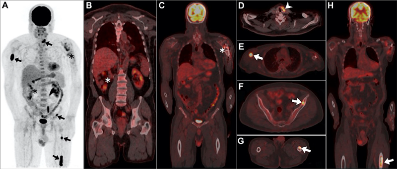
Figure 1
Figure 1. A 68-year-old male patient was recently diagnosed with renal cell carcinoma (RCC) of the right kidney. This was established after a major complaint of left arm pain, which necessitated further evaluation by pan-computed tomography (CT), which revealed evidence of a primary right renal malignancy and a single metastatic destructive left humeral shaft lesion. There was no radiological evidence of any other lesions elsewhere. Therefore, surgical interventions for both the primary and metastatic lesions were performed and included right nephrectomy and left humeral metastectomy with open reduction and internal fixation. The patient was then transferred to our cancer center for further management. Before starting therapy, a multidisciplinary clinic evaluation advocated further evaluation with 18F-fluorodeoxyglucose positron emission tomography/CT (18F-FDG PET/CT) before therapy initiation. The scan was conducted following the standard protocol after 60 min of intravenous administration of 7.3 mCi of 18F-FDG. Maximum intensity projection image revealing evidence of multiple hypermetabolic metastatic extrarenal sites (A; arrows). This was observed concurrently with unremarkable surgical sites for both the primary tumor and the previously identified metastatic left arm lesion (A-C; asterisks). Notably, there is evidence of an intense hypermetabolic focus involving the left thyroid cartilage (maximum standardized uptake value of 13.9; arrowhead, D). In addition, multiple hypermetabolic intramedullary lesions were observed within the right proximal humerus, left iliac bone, and left femoral shaft (arrows; E-H). Histopathological examination of the oligometastatic lesions was considered unnecessary because of the patient’s refusal and its lack of impact on the forthcoming management strategy. The hypermetabolic left thyroid cartilage deposit, which lacks identifiable structural characteristics, strongly suggests metastasis rather than primary involvement (1). This inference is supported by its distinct 18F-FDG uptake, which is contrary to typical primary cartilaginous lesions, which usually exhibit lower 18F-FDG uptake and evident radiographic manifestations (2). The lesion’s unifocal and intense 18F-FDG expression further diminishes the likelihood of a benign process. RCC is a lethal tumor that accounts for approximately 3% of all adult malignancies (3). The adoption and widespread use of molecular imaging techniques have led to a noteworthy rise in the accidental discovery of atypical metastatic manifestations (4). Our patient was initially diagnosed with a primary renal malignancy based on the presence of left arm discomfort caused by metastasis. Therefore, the traditional belief that primary RCC signs and symptoms should precede metastatic manifestations is more of an exception to a widespread principle. The progression of metastatic RCC does not always adhere to the traditional sequential metastatic pattern, suggesting a complex metastatic pathway (5). To date, numerous uncommon sites of metastasis have been identified (5). To the best of our knowledge, this is the first imaging instance where a patient with renal RCC established thyroid cartilage deposition as depicted on 18F-FDG PET/CT. Thyroid cartilage metastasis is rare in all types of malignancies. Poor vascular supply of cartilaginous tissue is considered a contributing factor to its reduced tendency for metastasis (1). Nonetheless, there have been only a small number of documented instances where metastasis has occurred in the thyroid cartilage, typically associated with advanced cancer and serving as an indication of widespread metastatic disease (1,6,7,8,9). What makes this case more interesting is that thyroid cartilage metastasis is observed in patients with oligometastases. Notably, the previous evaluation using pan CT failed to depict cartilage and bone marrow oligometastases. This signifies the vital utility of 18F-FDG PET/CT in the evaluation of RCC disease for baseline staging and beyond.
View larger Download

Footnotes

Ethics

Informed Consent: An informed consent was obtained from the patient.

Authorship Contributions

Surgical and Medical Practices: A.A-I., A.S.A., Concept: A.A-I., A.S.A., B.A., A.A., Design: A.A-I., A.S.A., B.A., K.A-R., Data Collection or Processing: A.A-I., A.A., Analysis or Interpretation: A.A-I., Literature Search: A.S.A., Writing: A.A-I., A.S.A., B.A., A.A., K.A-R.

Conflict of Interest: No conflicts of interest were declared by the authors.

Financial Disclosure: The authors declare that this study has received no financial support.

References

1

Gaber AM, Núñez R, Delpassand E. Cases of thyroid cartilage metastasis as abnormal findings seen in prostate cancer patients visualized by 68Ga-PSMA-11 PET/CT. Egypt J Radiol Nucl Med. 2021;52:134.

2

Mendoza H, Nosov A, Pandit-Taskar N. Molecular imaging of sarcomas with FDG PET. Skeletal Radiol. 2023;52:461-475.

3

Siegel RL, Miller KD, Wagle NS, Jemal A. Cancer statistics, 2023. CA Cancer J Clin. 2023;73:17-48.

4

Griffin N, Gore ME, Sohaib SA. Imaging in metastatic renal cell carcinoma. AJR Am J Roentgenol. 2007;189:360-370.

5

Sountoulides P, Metaxa L, Cindolo L. Atypical presentations and rare metastatic sites of renal cell carcinoma: a review of case reports. J Med Case Rep. 2011;5:429.

6

Tripathy S, Parida GK, Singhal A, Shamim SA, Kumar R. Thyroid Cartilage Metastases on F-18 Fluorodeoxyglucose Positron Emission Tomography-Computed Tomography: A Tale of Two Cases with a Brief Review of Literature. Indian J Nucl Med. 2019;34:220-222.

7

Gupta N, Verma R, Belho ES. Metastatic Thyroid Cartilage Lesion from Prostatic Adenocarcinoma onGa-Prostate-Specific Membrane Antigen Positron Emission Tomography-Computed Tomography Scan: Case Series. Indian J Nucl Med. 2021;36:183-8.

8

Tupalli A, Damle NA, Thankarajan AS, Mangu BS, Kumar A, Khan D, Sagar S, Bal C. An Unusual Case of Simultaneous Cricoid and Thyroid Cartilage Metastases from Prostatic Adenocarcinoma onGa-PSMA PET/CT. Nucl Med Mol Imaging. 2020;54:61-62.

9

Usmani S, Ahmed N, Ilyas MW, Murad S, Al Kandari F. Rare Thyroid Cartilage Metastasis From Breast Cancer Visualized on 18F-NaF PET/CT. Clin Nucl Med. 2021;46:43-44.

