

Abstract
Neurolymphomatosis (NL) is a rare clinical condition characterized by the infiltration of malignant lymphocytes into the cranial or peripheral nerves, nerve roots, or plexus. Diagnosis can be clinically challenging due to its variable presentation. It usually occurs in B cell lymphoma; however, a few cases of extranodal killer/T cell lymphoma. Most cases present at a secondary site in patients with primary site in remission. 18Fluorine fluorodeoxyglucose positron emission tomography/computed tomography plays an important role in the early detection of NL, resulting in timely treatment. We present a case of a 24-year-old male with nasal natural killer T cell lymphoma who initially responded to treatment but relapsed with NL based on clinical and radiological findings.
Keywords: Neurolymphomatosis, natural killer T cell lymphoma, 18Fluorine fluorodeoxyglucose positron emission tomography/computed tomography, lymphom
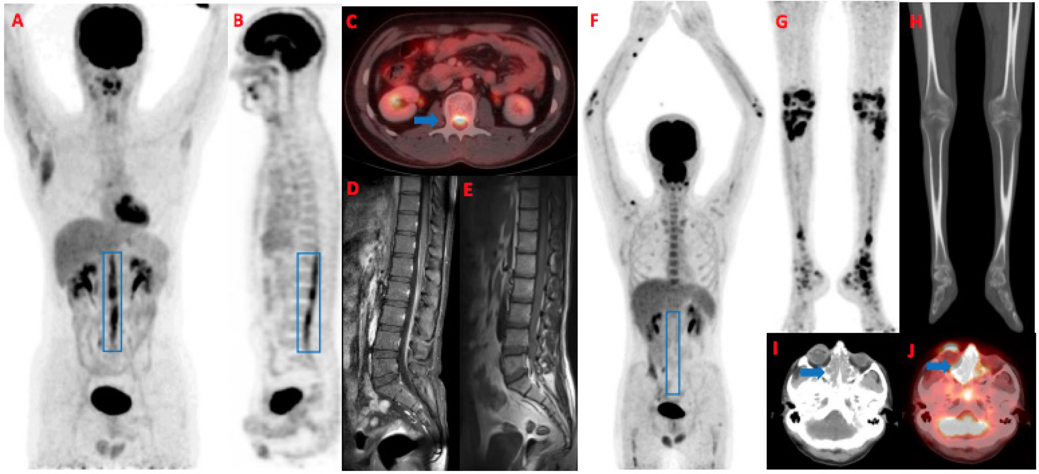
NL is a rare condition. Diagnosis can be clinically challenging due to its variable presentation. Literature review shows that, most commonly, it is associated with non-hodgkin lymphoma (,). Approximately 80% of the cases are associated with B cell lymphoma compared with T cell lymphoma. Among T cell lymphomas, NKTL is a rare entity that occurs at unusual sites, such as the central nervous system, skin, and nasopharynx. Despite aggressive treatment, it usually follows a rapidly progressive course (). It is important to differentiate NL from non-cancerous conditions, such as the Miller Fischer variant of Guillain-Barre Syndrome, inflammatory radiculopathy, neuropathies, and chemoradiotherapy-induced damage to the nerves. That history proves to be of paramount importance in making definitive diagnosis. Although histopathology remains the gold standard for diagnosing NL, due to its limitations, MRI and PET-CT play a major role in the diagnosis of NL, with sensitivity of 87.5% and 100%, respectively (,,). Biopsy is associated with an increased risk of permanent nerve damage, and it is usually reserved for cases that are not diagnosed on history and imaging. Within our institution, over a span of the last 15 years, there are 9 reported cases of NL on 18F-FDG PET/CT (,), of which only our patient was of NKTL. Therefore, clinicians reporting PET scans should be cognizant of the fact that NL is likely to represent the aggressive extra-nodal lymphoma spectrum.
Footnotes
Ethics
Informed Consent: An informed consent was obtained from the patient.
Authorship Contributions: Concept: S.M.G., P.A.A.A.Q., Design: S.M.G., A.H., P.A.A.A.Q., H.B., Data Collection or Processing: S.M.G., P.A.A.A.Q., Analysis or Interpretation: S.M.G., A.H., P.A.A.A.Q., H.B., Literature Search: S.M.G., Writing: S.M.G., A.H., P.A.A.A.Q.
Conflict of Interest: No conflicts of interest were declared by the authors.
Financial Disclosure: The authors declare that this study has received no financial support.
References
Pan Q, Luo Y. Recurrence of nasal type NK/T cell lymphoma presenting as neurolymphomatosis on 18F-FDG PET/CT: a case report and literature review. Medicine (Baltimore). 2020; 99:e18640.
DeVries AH, Howe BM, Spinner RJ, Broski SM. B-cell peripheral neurolymphomatosis: MRI andF-FDG PET/CT imaging characteristics. Skeletal Radiol. 2019; 48:1043-1050.
Lee J, Suh C, Park YH, Ko YH, Bang SM, Lee JH, Lee DH, Huh J, Oh SY, Kwon HC, Kim HJ, Lee SI, Kim JH, Park J, Oh SJ, Kim K, Jung C, Park K, Kim WS. Extranodal natural killer T-cell lymphoma, nasal-type: a prognostic model from a retrospective multicenter study. J Clin Oncol. 2006; 24:612-618.
Choi YJ, Shin JA, Kim YH, Cha SJ, Cho JY, Kang SH, Yi SY, Lee HR. Neurolymphomatosis of brachial plexus in patients with non-hodgkin’s lymphoma. Case Rep Oncol Med. 2013; 2013:492329.
Baehring JM, Batchelor TT. Diagnosis and management of neurolymphomatosis. Cancer J. 2012; 18:463-468.
Soni N, Ora M. FDG PET/CT in neurolymphomatosis. Radiology. 2021; 298:36.
Yaseen MT, Aziz PAA, Siddique K, Tariq TA. Neurolymphomatosis of the sciatic and tibial nerves. J Coll Physicians Surg Pak. 2019; 29:S86-S88.
Riaz S, Naz F, Bashir H, Niazi IK. “Bottle brush sign”- Spinal meningeal disease on 18F-FDG PET-CT scan. Clin Nucl Med. 2016; 41:726-727.

